On-Pump Beating Heart Complete Arterial Coronary Revascularization in a Patient with Porcelain Aorta

Francisco J. Vázquez Roque, Yuri Medrano Planas, Ernesto Chaljub Bravo, Luís A. González Borges and Yolepsis Quíntero Fleites

Department of Cardiovascular Surgery. Heart Center in Santa Clara, Villa Clara, Cuba

Academic Editor: Shahzad Gull Raja

Cite this Article as:

Francisco J. Vázquez Roque, Yuri Medrano Planas, Ernesto Chaljub Bravo, Luís A. González Borges and Yolepsis Quíntero Fleites (2014), "On-Pump Beating Heart Complete Arterial Coronary Revascularization in a Patient with Porcelain Aorta," Journal of Research in Cardiology, Vol. 2014 (2014), Article ID 129108,
DOI: 10.5171/2014.129108

Copyright © 2014. Francisco J. Vázquez Roque, Yuri Medrano Planas, Ernesto Chaljub Bravo, Luís A. González Borges and Yolepsis Quíntero Fleites. Distributed under Creative Commons CC-BY 3.0

Abstract

Extensive calcification of the ascending aorta is an infrequent but alarming finding while performing coronary artery bypass grafting surgery. Porcelain aorta (PA) requires adjustments in cardiopulmonary bypass as well as in revascularization techniques. Our case consists of a 64 years old woman with left main stenosis plus  three vessels coronary artery disease and PA in whom on-pump beating heart total arterial myocardial revascularization using both internal mammary arteries as a composite T graft was performed with good outcomes at short and middle terms.

Keywords: Coronary artery bypass grafts, CABG, CABG, arterial grafts, Aorta/aortic.

Introduction

A severely calcified aorta carries a high risk of atheroembolism and bleeding during cardiac surgery. PA has an incidence of 14% to 29% in patients with coronary artery disease combined with aortic valve stenosis according to a paper by Hokenek et al., (2011). A reason for this wide range is the varying definition of PA. Imaging methods to identify PA preoperatively are chest roentgenography, computed tomography, and cardiac catheterization. Sometimes a heavily calcified aorta can remain undetected until palpation after sternotomy. Fatal stroke and profuse bleeding during aortic clamping and cannulation may be caused by crushing the calcified aortic layers. Embolism of mobile atheromathous fragments into the systemic circulation can also occur. Most of these patients present numerous additional risk factors (advanced age, coronary artery disease, carotid artery stenosis, history of stroke and peripheral arterial disease). Thus, this population represents a truly high-risk cohort. Various techniques are described to avoid aortic cannulation and clamping known as aortic no-touch techniques as those studied by Emmert et al., (2012) and Kumar et al., (2011). When PA patients need coronary artery bypass grafting, the no-touch beating heart technique seems to be nowadays the best alternative.

Case report

We present a 64-year-old woman was admitted with progressive angina, Canadian Cardiovascular Society Class III at admission, not relieved by optimal medical therapy. Antecedents of arterial hypertension, diabetes, and hypercholesterolemia as risk factors for coronary artery disease were present. Preoperative chest X-ray (fig. 1a) showed severe ascending aortic calcification, which was also visible during cardiac catheterization (Fig. 1b). Significant left main coronary artery stenosis and significant proximal lesions of the left anterior descending artery (LDA), left circumflex artery, and the right coronary artery (Fig. 1c y d) demanded surgical revascularization.

129108-fig-1

Figure 1: Porcelain aorta and significant left main coronary artery stenosis plus severe three vessel diseases
a: chest X-ray.
b: Cardiac catheterization.
c: Significant left main coronary artery stenosis.
d: Significant lesions in right coronary artery.
 

Procedure description

We decided to undertake total arterial myocardial revascularization with both internal thoracic arteries. After a median sternotomy, the pericardial cavity was entered. Intraoperative examination revealed that the entire wall of the ascending aorta was severely calcified, thus, unsuitable neither for arterial cannulation nor for side clamping. Both internal thoracic arteries (ITAs) were dissected along their entire length in a skeletonized fashion with opening of both pleuras and using low intensity electrocauterization. The left side of the opened pericardial sac was incised down vertically just opposite the pulmonary artery until 2 cm above the phrenic nerve. The left ITA (LITA) was divided proximal the distal bifurcation. LITA flow was over 100 ml per minute. Afterward, its distal end was occluded with a soft bulldog clamp and covered with a papaverine solution impregnated gauze pad. Then, the right ITA (RITA) was transected at its origin from the subclavian artery and distally at the level of its bifurcation. It was anastomosed in a T fashion to LITA (Fig. 2 a) at the level of the pulmonary valve. Both internal thoracic arteries were preserved in papaverine impregnated gauze. Three deep traction pericardial sutures in the left pericardial sac were placed using 3-0 Prolene (Ethicon, Somerville, NJ) to accomplish adequate exposure of the coronary vessels. Off-pump beating coronary artery bypass grafting was intended, but deterioration of the patient’s hemodynamic status (non-sustained ventricular tachycardia and hypotension during cardiac repositioning) demanded cardiopulmonary circulatory support. Timely insertion of cannulas thru the right femoral artery and the right atrial appendage for arterial perfusion and venous drainage respectively were performed. Peripheral arterial cannulation was considered mandatory given the overwhelming risk of aortic manipulation in cases of PA. Furthermore, preoperative ultrasonographic evaluation of limbs and carotid arteries in our patient revealed only mild atheromatous affection of the femoral arteries. Subclavian arterial cannulation is an alternative approach which, despite its proximity to the surgical field, requires further time to anastomose an interposition graft for the cannula. Partial cardiopulmonary bypass was started. Then we evaluated if the right AMI was long enough to reach the posterior descending coronary artery (PDA) before emptying the heart. Full flow cardiopulmonary bypass at 36°C was established. The heart remained beating to avoid any clamping procedure involving the friable aorta, either external or endoluminal, necessary for successful cardioplegia. Pledgeted stiches with 3/0 prolene surrounding epicardial coronary arteries placed on tourniquets were used to occlude the target vessels proximal to the anastomotic sites. Octopus 4 cardiac suction stabilizer was employed. For better exposure of the lateral and inferior left ventricular wall we used a Starfish stabilizer.


129108-fig-2
 
Figure 2: Complete arterial myocardial revascularization procedures 
a: RIMA is anastomosed in a T fashion to the pedicled LITA.
b: LIMA anastomosed to LDA.
c y d: Sequential anastomosis were performed to the OMA  and PDA with RITA.

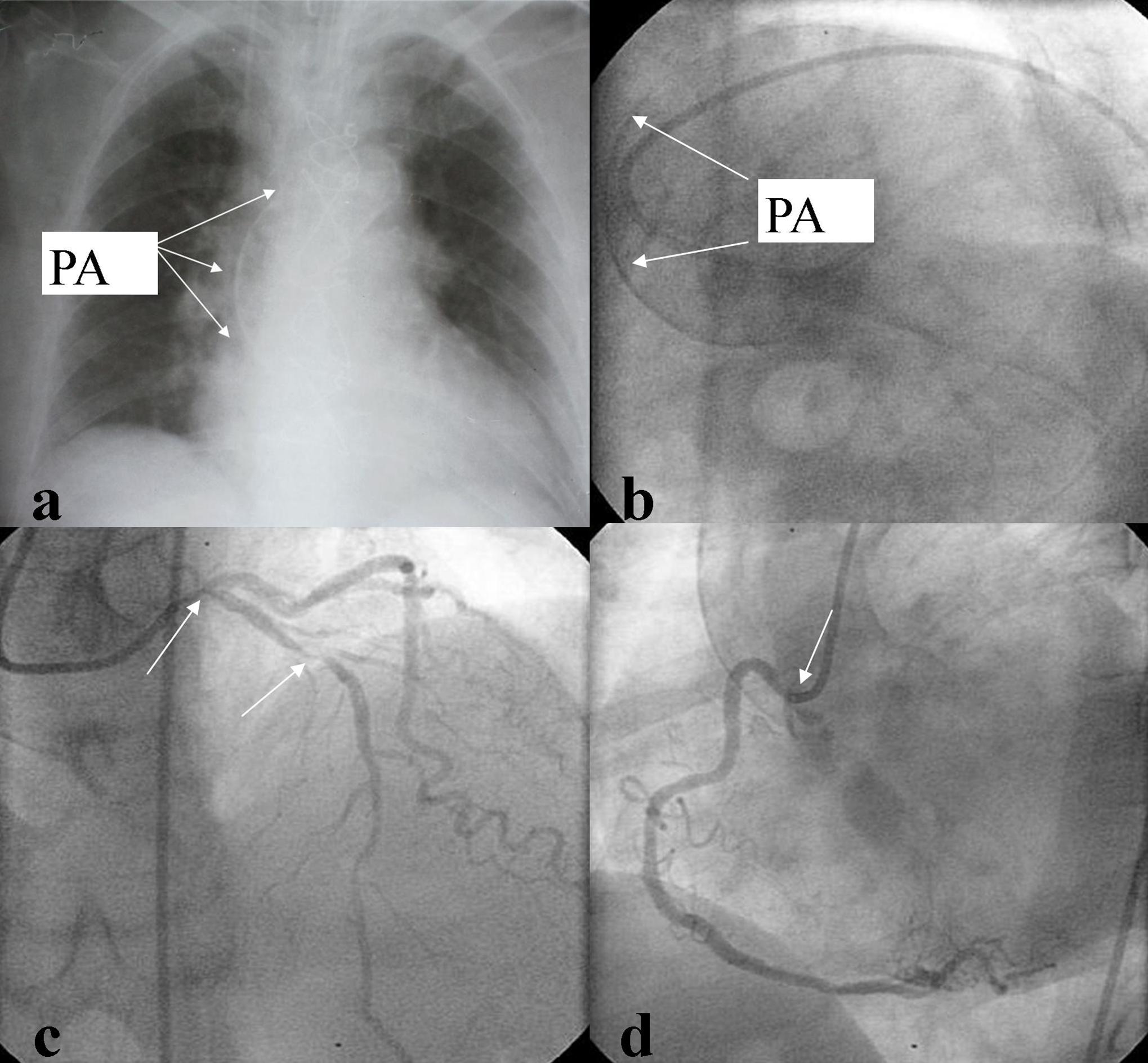
First, we grafted the pedicled LITA to the LAD with an end-to-side anastomosis (Fig. 2 b). Then obtuse marginal branch (OM), and PDA of RCA were anastomosed sequentially with RITA side-to-side and end-to-side respectively (Fig. 2 c y d). All anastomoses were performed with 8-0 Prolenesuture. Rest of the surgical procedure was completed in the usual fashion without complications. Duration of extracorporeal circulation was 83 minutes. Postoperative course and convalescence progressed uneventfully. After ten days of in hospital postoperative care the patient was discharged with evident clinical and hemodynamic recovery. Eighteen months after hospital discharged he was asymptomatic (New York Heart Association Class I, Canadian Cardiovascular Society Class I). Transthoracic echocardiography showed normal function and size of both ventricles. Postoperative multi-slice computed tomographic angiography showed patency of every graft.

Comments

Saphenous vein graft atherosclerosis continues to be the major cause of coronary artery bypass grafting late failure. The use of both internal thoracic arteries for coronary revascularization has been demonstrated to be advantageous over the use of single LIMA in combination with vein grafts with respect to survival, freedom from angina and repeated revascularization procedures. Complete arterial myocardial revascularization is the procedure of choice in young adults and in patients with porcelain aorta. Benefits of complete arterial revascularization are well established as reported in the research by Fiori et al., (1990). When employing composite grafts, arterial flow to the revascularized left ventricular myocardium can be provided exclusively by in-situ LITA. In our patient the LITA flow was over 100 ml per minute. Semiskeletonized, and skeletonized techniques harvesting internal thoracic arteries result in longer arterial conduits which can reach the lateral and posterior wall of the heart, such as in our case. Sometimes, RITA is too short for grafting coronary arteries in the posterior wall of the heart. Alternatively, when the LAD lesion is proximal, a transected short segment of distal LIMA could be anastomosed to the distal RITA to accomplish complete revascularization. We have used this technique in several patients with good results. A short segment of the radial artery or saphenous vein may be used as well as disclosed by a paper by Davierwala (2013).

We consider off-pump beating heart coronary artery surgery as the best choice in PA patients with hemodynamic stability meanwhile in patient without hemodynamic stability we prefer on-pump beating heart revascularization with a combination of peripheral arterial cannulation and central venous drainage for cardiopulmonary bypass.

References

1. Davierwala, P. M. (2013) “No-touch aorta off-pump coronary bypass operation: arteriovenous composite grafts may be used as a last resort,” Annals of Thoracic Surgery, 95 (3) 846-852.
PublisherGoogle Scholar

2. Fiore, A. C, Naunheim, K. S, Dean,  P.(1990) “Results of internal thoracic artery grafting over 15 years: single versus double grafts,” Annals of  Thoracic Surgery, 49:202—209.
PublisherGoogle Scholar

3.    Hokenek, F, Gursoy, M, Ozgol, I, Gulcan, F. and Kınoglu, B. (2011) “A new proximal anastomosis technique in two patients with severely calcified aorta,” Annals of   Thoracic Surgery, 92 (5)1914—1916.
PublisherGoogle Scholar

4.  Emmert, M. Y, Seifert, B, Wilhelm, M, Grünenfelder, J, Falk, V, Salzberg, S. P. (2012) “Aortic no-touch technique makes the difference in off-pump coronary artery bypass grafting,”Journal of Thoracic and Cardiovascular  Surgery, 143 (4) 995. 

5.  Kumar, K, Deval, M, Manoria, P, Chand, P. and Jagdale, L. (2011) “Off pump CABG with aortic-no-touch using bilateral in-situ IMA: a new technique,” Indian Journal of  Thoracic and  Cardiovascular Surgery  27 (4)165—168.
PublisherGoogle Scholar 

Shares