Temporal Relation of Myocardial Ischemia to Nocturnal Desaturation of Oxygen in Patients with Coronary Artery Disease

Dette FG1, Cassel W2, Penzel T3, Koehler U4 and  Graf J5

1 Department of Anaesthesiology, University Medical Center of the Johannes Gutenberg-University Mainz, Langenbeckstraße Mainz, Germany

2,4 Department of Internal Medicine — Pneumology and Center of Sleep Medicine, Philipps-University Marburg, Baldinger Straße, Marburg, Germany

3 Center of Sleep Medicine, Charité-University Medical Center Berlin, Charitestraße Berlin, Germany

5 Klinikum Stuttgart, Kriegsbergstraße Stuttgart, Germany

Academic Editor: Harald H. Schäfer

Cite this Article as:

Dette FG, Cassel W, Penzel T, Koehler U and Graf J (2014), " Temporal Relation of Myocardial Ischemia to Nocturnal Desaturation of Oxygen in Patients with Coronary Artery Disease", Journal of Research in Cardiology, Vol. 2014 (2014), Article ID 313309, DOI: 10.5171/2014.313309

Copyright © 2014. Dette FG, Cassel W, Penzel T, Koehler U and Graf J. Distributed under Creative Commons CC-BY 3.0

Abstract

Purpose: Obstructive sleep apnea (OSA) is frequently associated with coronary artery disease. Causal relation of nocturnal respiratory disorders to the onset of myocardial ischemia has been discussed controversially. Methods:Patients with coronary artery disease (CAD) were enrolled in the present pilot study. Overnight polygraphy and 12-lead ECG were performed in all patients. Patients with nocturnal myocardial ischemia in the study night were eligible for analysis of breathing parameters preceding myocardial ischemia. Backwards from the first second of myocardial ischemia, a preceding period of 900 seconds was analysed for OSA and subsequent reduction of oxygen saturation. The frequency of respiratory events within this period was compared to frequency within the total time of sleep assessment.Results: We present data of 19 patients (4 women; 15 men) aged 45 to 79 years (mean 64.1 years; ± 9.4 (standard deviation)) with a mean body mass index (BMI) of 29.5 kg(m2)-1 ± 4.4. No patient had been diagnosed with OSA before study participation. In all patients an Apnea-Hypopnea-Index (AHI) 5/h (median 11/h; IQR: 8/25), in 9 patients (47%) an AHI of 15/h (median 25; IQR: 20/47.5) was calculated. Nocturnal myocardial ischemia was found in 8 patients and furthermore, one patient reported nocturnal chest pain. Preceding myocardial ischemia OSA was more frequent compared to the time without ischemia 100% (95%/100%) versus 66.8% (58%/75.9%); p= 0.012. Conclusion: The present study concludes that nocturnal myocardial ischemic events frequently occur with a temporal related accumulation of preceding OSA.

Keywords: Myocardial ischemia; nocturnal hypoxemia, OSA, temporal relation

Introduction

Strong association between sleep disordered breathing (SDB) and, in particular, between obstructive sleep apnea (OSA) and cardiovascular disease has been highlighted in previous studies by Shahar et al. (2001) and Franklin and co-workers (1995). Besides others, OSA has been associated with ischemic heart disease and hypertension as shown by Peker and colleagues (2002). OSA has shown to be a risk factor for coronary events and a predictor for cardiovascular death found by Shah et al. (2010) and Martinez et al. (2012). Causal relation of OSA with regard to the onset of nocturnal myocardial ischemia has been controversially discussed by Keyl et al. (1994), Smith et al. (1996) and Gögenur et al. (2004). OSA is characterized by recurrent occlusion of the upper airway with subsequent total or partial flow limitation, as well as intermittent hypoxemia. Normally, this situation is terminated by an arousal reaction with a sympathetic burst. Blood pressure and heart frequency increase within a hypoxemic condition. We hypothesized intermittent hypoxemia to be a trigger for nocturnal myocardial ischemia prior to elective surgery.

This study was designed to analyse the temporal association of myocardial ischemia detected with nocturnal 12-lead-Holter-electrocardiogram (ECG) and related parameters indicative for OSA.

Methods

The study protocol was approved by the ethics committee of the Philipps-University of Marburg (AZ 76/04). Informed written consent was obtained from all patients.

Coronary artery disease (CAD) was pre-known in all patients who came routinely for check-up after previous coronary intervention (coronary stenting) to the hospital. Stress ECG was performed in all patients, and those with pathological result were scheduled for coronary angiography. These patients were eligible for study enrolment, and overnight polygraphy with time-synchronized 12-lead Holter-ECG was performed the night before. Patient`s coronary status was documented according to results of coronary angiography. Re-stenosis ≥ 70% of minimum one coronary vessel was found in all assessed patients.

Patients who had a history of drug or alcohol abuse and patients with a long term sleep medication were excluded. Furthermore, patients were excluded if they had abnormalities in the ECG such as left and right bundle block. Nocturnal breathing assessment was made by overnight polygraphy (SOMNOcheck®, Weinmann, Germany) including assessment of peripheral arterial oxygen saturation, breathing flow, snoring and movement of thorax and abdomen. This assessment was performed from 10 p.m. to 6 a.m. on the cardiology ward in rooms with two beds. Periods with reduction of breathing flow of more than 30% of baseline with an accompanying decrease of oxygen saturation of ≥4% of baseline were scored as hypopneas; periods with a total flow-suppression of more than 10 seconds were counted as apneas. Apneas and hypopneas were manually assessed. Patients received no sedative agents the evening before measurement. ECG monitoring was performed using a 12-lead-recorder (WelchAllyn CardioPerfect Holter-ECG). The ST-segment was measured 0.08 s after the J-point. Ischemia was defined by ST changes in two contiguous leads. First, it was planned to assess data of ECG with a related commercial computer algorithm. Unfortunately the frequency of false results and artefacts via automated assessment was high. Therefore, data were manually analyzed by an experienced physician blinded with respect to medical records and the individual patient. These results were taken for final analysis. Over 24 hours, time on Holter-ECG differed from time on SOMNOcheck device a minimum of 2 seconds to a maximum of 8 seconds. Both devices were synchronized before each recording and equipped with a new battery before starting.

Previous data of other authors (Mooe et al. (2000) and Reeder et al. (1991)) show temporal relationship between ST-depression and decrease of oxygen saturation within a period of 2 minutes and 30 minutes, respectively. In the present study a period for time assessment of 15 minutes was chosen. For the final assessment, data were analysed in segments of seconds. Backwards from the first second of myocardial ischemia on ECG a preceding time of 900 seconds (15 minutes) was analysed for apneas, hypopneas and oxygen desaturations in the same individual. Results were set in relation to the frequency of apneas and hypopneas within the total time of sleep assessment (10 p.m. to 6 a.m.).

Statistical Analysis

Statistical analysis was performed by a statistician blinded to the individual cases using SPSS (version 15.0; SPSS Inc., Chicago, Illinois). Data were checked for deviations from the normal distribution by means of the Kolmogorov-Smirnov one sample test. Apnoea-Hypopnoea-Index (AHI) and arterial oxygen saturation were not normally distributed. Thus, a non-parametric test (Wilcoxon signed rank test) was applied. Results are given as median, first and third quartile. Results of normally distributed data are presented as mean and standard deviation (SD). Differences of normally distributed data were assessed by Student`s t-test. A p-value < 0.05 was considered statistically significant.

Results

Within the present study 22 patients were prospectively recruited. Agreement was revoked prior to polygraphy by two patients, and data quality was poor in one case. These data sets were therefore discarded. Thus, we are presenting data of 19 patients (4 women and 15 men) aged 45 to 79 years (mean 64.1 ± 9.4 years) with a mean body mass index (BMI) of 29.5 ± 4.4 kg (m2)-1. Patient´s characteristics are shown in Table1. No patient had a previous diagnosis of OSA.

In all patients, an AHI ≥ 5/h was observed (median 11/h; IQR: 8/25) and in 9 of 19 patients (47%) an AHI of ≥ 15/h (median 25/h; IQR: 20/47.5).

Nocturnal reduction of oxygen saturation was frequently found in all patients, whereas overall 13 episodes of nocturnal myocardial ischemia were detected only in 8 of 19 patients (42%). Both, ST depression and ST elevation were considered to be ST abnormalities. However, only ST depression occurred in the study population. One of these patients reported nocturnal chest pain the morning after measurement. A cutout of the nocturnal ECG of a study patient is shown in Figure1. Patients with myocardial ischemia and patients without did not differ with regard to the characteristics shown in Table1 Table1: Characteristics of the study population. (BMI = body mass index [kg (m2)-1]; SpO2 (n) = nadir arterial oxygen saturation; SpO2 (oa) = arterial oxygen saturation on average; AHI = Apnoea-Hypopnoea-Index; MI = number of detected myocardial ischemic events)

Table 1: Characteristics of the study population. (BMI = body mass index [kg (m2)-1]; SpO2 (n) = nadir arterial oxygen saturation; SpO2 (oa) = arterial oxygen saturation on average; AHI = Apnoea-Hypopnoea-Index; MI = number of detected myocardial ischemic events)

 
313309-tab-1
313309-fig-1
 
Figure 1: Shown is a cutout of the nocturnal 12-lead ECG of a study patient who reported chest pain the morning after within a period of myocardial ischemia
 

(age: p=0.59; BMI: p=0.46; nadir SpO2: p=0.9; SpO2 on average: p=0.53; AHI: p=0.45; number of stenosed coronary vessels: p=0.44). Co-morbidity profile and patients’ medication is shown in Table2. Episodes of nocturnal hypoxemia were also frequently detected without temporal related ischemic events in ECG. To quantify these results, data of patients with nocturnal ischemic events in ECG (8 of 19) were finally analyzed in segments of seconds. Moreover, the distribution of breathing events and ischemic events in ECG were analyzed over the time of the study night. This analysis yielded to the following results:

Over a median of 167 seconds (IQR: 76/279 seconds) myocardial ischemia was detected. Within the preceding 900 seconds, OSA was found with a frequency of 100% (95%/100%). Within the time without ischemic events, OSA occurred only in 66.8% (58%/75.9%); p= 0.012 (Figure 2).   

Table 2: Co-morbid profiles and medication of the study participants (ACE-I = angiotensin-converting enzyme inhibitors; CG= cardiac glycosides; AT1-RA= AT1-receptor antagonists; HLP= hyperlipoproteinemia; Smoker= smoker and former smoker)
 
313309-tab-2
 
313309-fig-2
Figure 2: Frequency of sleep disordered breathing the time before myocardial ischemia (left) and the time without (right)
  

Discussion

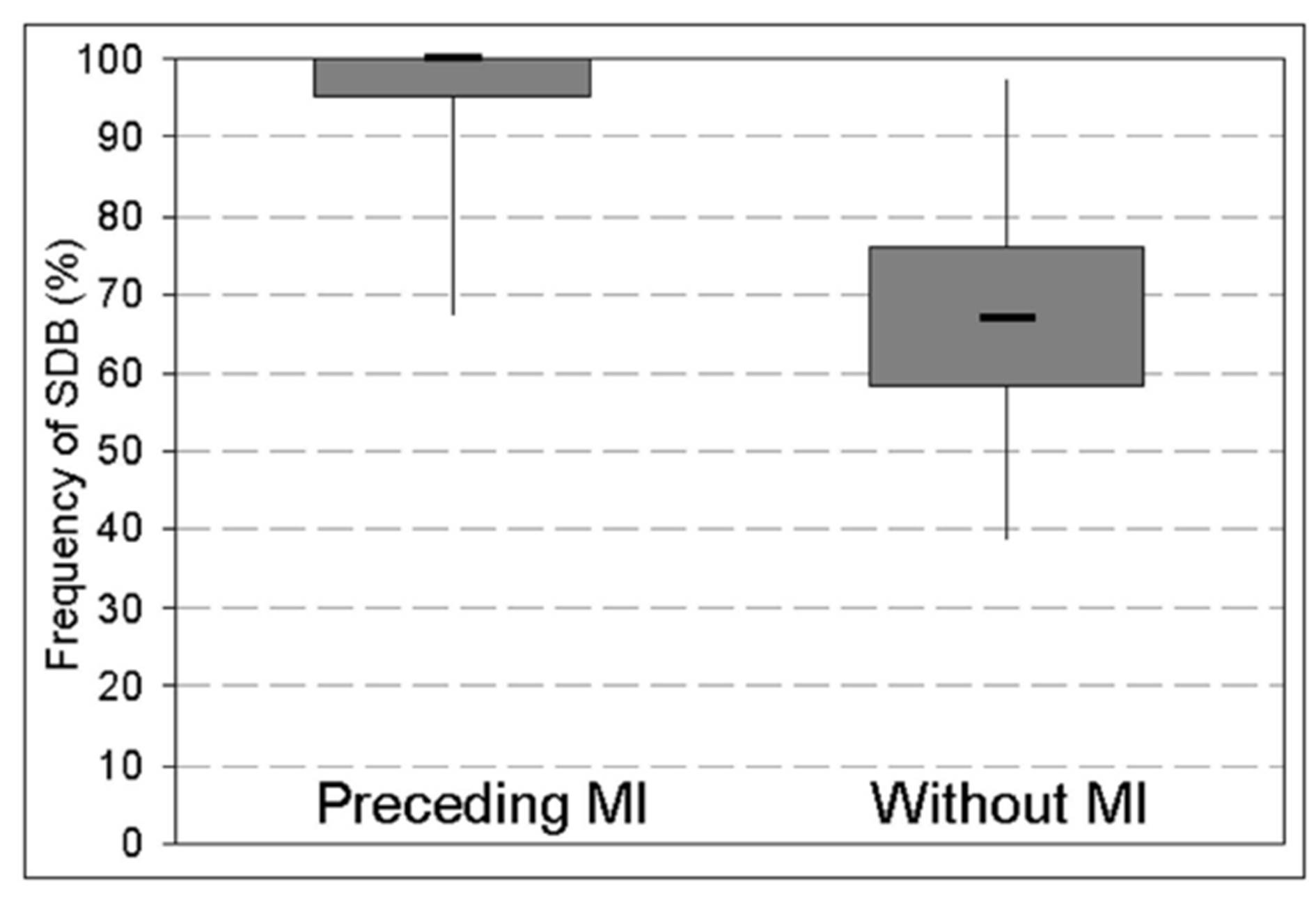
The present results indicate a high frequency of previously unrecognized and therefore untreated OSA in a group of patients with CAD. An AHI of ≥ 5/h with consecutive reduction of arterial oxygen saturation was observed in all recruited patients. Of note, all patients with myocardial ischemia during the observation period experienced OSA in the preceding period of 900 seconds.

A high prevalence of OSA in patients with CAD has been previously demonstrated by Shahar et al. (2001) and Peker et al. (2002). Marin and co-workers (2005) showed higher incidence of cardiovascular events in patients with OSA in comparison to patients without. Furthermore, SDB was associated with re-stenosis, remodeling (Steiner et al. 2008) and cardiac mortality after percutaneous intervention as shown by Yumino et al. (2007). Treatment of SDB with continuous positive airway pressure (CPAP) was presumed by Drager et al. (2007) to impede progression of atherosclerosis to clinically important cardiovascular diseases. Milleron and colleagues (2004) showed a reduction of new cardiovascular events in patients with CAD when sleep disordered breathing had been treated by CPAP-therapy. On the other hand, Shah and colleagues (2013) found OSA to have a protective role in acute myocardial infarction by chronic but mild intermittent hypoxic preconditioning.

In the present study, a temporal association of nocturnal respiratory disorders on myocardial ischemia was hypothesized. As described by other authors, nocturnal impairment of oxygen saturation occurred frequently, whereas myocardial ischemia was only detected in less than 50% of the study population during the study night. The presumed temporal association was controversial discussed in the literature. Keyl and colleagues (1994) as well as Smith and co-workers (1996) found no association between oxygen saturation and myocardial ischemia, whereas Reeder et al. (1991) described myocardial ischemia as temporally associated with hypoxemia. These results are in agreement with our findings, as accumulation of OSA events preceding episodes of myocardial ischemia was found.

Arbab-Zadeh and co-workers (2009) demonstrated the inability of diseased coronary artery segments to react to hypoxemia. However, all patients of the study population had diseased coronary arteries. Thus, other factors i.e. rapid-eye-movement (REM) and non-REM sleep should be considered. Kim and colleagues (2008) found severe myocardial ischemia in an animal model with stenosed coronary arteries during non-REM sleep. Nocturnal myocardial ischemia potentially occurs if oxygen partial pressure in blood has fallen below a critical level. This threshold probably differs between subjects. Temporal and accumulating SDB as shown in the present study would lead to reduced level of oxygen partial pressure.  

Limitations and Strength

The present study was not performed with polysomnography (PSG). Therefore, an allocation of myocardial ischemia to sleep stages was not possible. It should be mentioned that nocturnal ischemic events frequently occur in REM sleep as shown by Schäfer et al. (1997). Furthermore, heart rate as an important determinant of myocardial oxygen consumption was not assessed and analyzed.

Due to its small sample size, the power of the study is limited. However, to the best of our knowledge this is the first study that employed long-term 12-lead-ECG for the detection of nocturnal myocardial ischemia associated with cardio-respiratory polygraphy. Body position while measuring ST abnormalities was not addressed within the analysis.

The present study concludes that nocturnal myocardial ischemic events frequently occur with a temporal related accumulation of preceding SDB.

References

1. Arbab-Zadeh A, Levine BD, Trost JC, Lange RA, Keeley EC, Hillis LD, Cigarroa JE (2009) “The effect of acute hypoxemia on coronary arterial dimensions in patients with coronary artery disease,” Cardiology 113:149-54.
Publisher Google Scholar

2. Drager LF, Bortolotto LA, Figueiredo AC, Krieger EM, Lorenzi GF (2007) “Effects of continuous positive airway pressure on early signs of atherosclerosis in obstructive sleep apnea,” Am J Respir Crit Care Med 176:706-12.
Publisher Google Scholar

3. Franklin KA, Nilsson JB, Sahlin C, Näslund U (1995) “Sleep apnoea and nocturnal angina,” Lancet 345:1085-7.
PublisherGoogle Scholar

4. Gögenur I, Rosenberg-Adamsen S, Lie C, Carstensen M, Rasmussen V, Rosenberg J (2004)
“Relationship between nocturnal hypoxaemia, tachycardia and myocardial ischaemia after major abdominal surgery,” Br J Anaesth 93:333-8
Publisher Google Scholar

5. Keyl C, Lemberger P, Rödig G, Dambacher M, Frey A (1994) “Hypoxaemia and myocardial ischaemia on the night before coronary bypass surgery,” Br J Anaesth 73:157-61.
Publisher Google Scholar

6. Kim SJ, Kuklov A, Kehoe RF, Crystal GJ (2008) “Sleep-induced hypotension precipitates severe myocardial ischemia,” Sleep 31:1215-20.
Publisher – Google Scholar

7. Marin JM, Carrizo SJ, Vicente E, Agusti AG (2005) “Long-term cardiovascular outcomes in men with obstructive sleep apnea-hypopnoea with or without treatment with continuous positive airway pressure: an observational study,”Lancet 365:1046-53.
Publisher Google Scholar

8. Martinez D, Klein C, Rahmeier L, Pacheco da Silva R, Fiori CZ, Cassol CM, Goncalves SC, Goncalves Bos AJ (2012) “Sleep apnea is a stronger predictor for coronary heart disease than traditional risk factors,” Sleep Breath 16:695-701
Publisher Google Scholar

9. Milleron O, Pillière R, Foucher A, de Roquefeuil F, Aegerter P, Jondeau G, Raffestin BG, Dubourg O (2004)
“Benefits of obstructive sleep apnoea treatment in coronary artery disease: a long-term follow-up study,” Eur Heart J 25:728-34.
PublisherGoogle Scholar

10. Mooe T, Franklin KA, Wiklund U, Rabben T, Holmström K (2000) “Sleep-disordered breathing and myocardial ischemia in patients with coronary artery disease,” Chest 117:1597-602.
Publisher Google Scholar

11. Peker Y, Hedner J, Norum J, Kraiczi H, Carlson J (2002) “Increased incidence of cardiovascular disease in middle-aged men with obstructive sleep apnea: a 7-year follow-up,” Am J Respir Crit Care Med 166:159-65.

12. Reeder MK, Muir AD, Foëx P, Goldman MD, Loh L, Smart D (1991) “Postoperative myocardial ischaemia: temporal association with nocturnal hypoxaemia,” Br J Anaesth 67:626-31.
PublisherGoogle Scholar

13. Schäfer H, Koehler U, Ploch T, Peter JH. (1997) Sleep-related myocardial ischemia and sleep structure in patients with obstructive sleep apnea and coronary heartdisease. Chest. 1997;111:387-93.
Publisher Google Scholar

14. Shah N, Redline S, Yaggi KH, Wu R, Zhao CG, Ostfeld R, Menegus M, Tracy D, Brush E, Appel WD, Kaplan RC (2013) “Obstructive sleep apnea and acute myocardial infarction severity: ischemic preconditioning?” Sleep Breath 17:819-26
Publisher Google Scholar

15. Shah NA, Yaggi HK, Concato J, Mohsenin V (2010) “Obstructive sleep apnea as a risk factor for coronary events or cardiovascular death,” Sleep Breath 14:131-36
PublisherGoogle Scholar

16. Shahar E, Whitney CW, Redline S, Lee ET, Newman AB, Javier Nieto F, O’Connor GT, Boland LL, Schwartz JE, Samet JM (2001) “Sleep-disordered breathing and cardiovascular disease: cross-sectional results of the Sleep Heart Health Study,” Am J Respir Crit Care Med 163:19-25.
PublisherGoogle Scholar

17. Smith HL, Sapsford DJ, Delaney ME, Jones JG (1996) “The effect on the heart of hypoxaemia in patients with severe coronary artery disease,” Anaesthesia 51:211-8.
PublisherGoogle Scholar

18. Steiner S, Schueller PO, Hennersdorf MG, Behrendt D, Strauer BE (2008) “Impact of obstructive sleep apnea on the occurrence of restenosis after elective percutaneous coronary intervention in ischemic heart disease,” Respir Res 9:50.
PublisherGoogle Scholar

19. Yumino D, Tsurumi Y, Takagi A, Suzuki K, Kasanuki H (2007) “Impact of obstructive sleep apnea on clinical and angiographic outcomes following percutaneous coronary intervention in patients with acute coronary syndrome,”
Am J Cardiol 99:26-30.
PublisherGoogle Scholar

Shares