Correlation between Images for Diagnosis Techniques in Temporomandibular Disorders

Giambartolomei Luis Augusto1, 2, Scavone Mario1, Brunotto Mabel2, Ferraris María Elsa2 and Azcona Sergio1

1Team Health and Education with Science (ESECi). Córdoba. Argentina

2School of Dentistry. National University of Córdoba. Argentina

Academic Editor: Andrea Å prláková-Puková

Cite this Article as:

Giambartolomei Luis Augusto, Scavone Mario, Brunotto Mabel, Ferraris María Elsa and Azcona Sergio (2014), "Correlation between Images for Diagnosis Techniques in Temporomandibular Disorders ," Journal of Research and Practice in Dentistry, Vol. 2014 (2014), Article ID 507487, DOI: 10.5171/2014.507487

Copyright © 2014 Giambartolomei Luis Augusto, Scavone Mario, Brunotto Mabel, Ferraris María Elsa and Azcona Sergio. Distributed under Creative Commons CC-BY 3.0

Abstract

The purpose of this research is to conduct a comparative analysis between two imaging techniques complementary to clinical diagnosis: the orthopantomogram (OPG) and the magnetic resonance imaging (MRI). Materials and Methods: An analysis of orthopatomograms (OPG) and magnetic resonance imaging (MRI) of right and left TMJ was carried out in 84 females and 24 males (average age: 31). 70 TMJ (problem cases) with medial or lateral disc displacement (MDD or LDD) and 25 TMJ (control cases) with no disc displacement (WNDD) were studied. There was a correlation between the intra-articular bio-mechanical state of TMJ diagnosed by MRI, and the vertical mandibular asymmetry (VMA) shown in the OPG. The data obtained from both techniques were analyzed by Kendall’s W Test statistic value — coefficient of concordance 0,206, degree of freedom 1 and P ≤ 005. Results: There was no association between the vertical mandibular asymmetry (VMA) measured by the OPG and the disc displacement (DD) observed in the MRI. Conclusion: Based on the results gathered from the mandibular OPG and the MRI of the TMJ, it was not possible to establish a direct relation between the mandibular asymmetry and the internal biomechanical TMDs.
Keywords: TMJ, orthopantomogram, magnetic resonance imaging, correlation, and asymmetry.

Introduction

The anatomical modification of the temporomandibular joint (TMJ) is one of the factors to be considered within TMD etiology. Giambartolomei (2003), Habets et al. (1998).There was a large vertical height condylar asymmetry in patients with TMD, compared to people from a control case group measured by panoramic radiograph called orthopantomogram (OPG). Kopp et al. (1979). As a replacement of that radiograph, the cross-sectional tomography is a further option for the diagnosis Pasquet et al, (2009) but it does not show the soft tissues. However, the OPG has proved to be extremely useful for complete luxation of the mandibular condyle cases. Anantharam (2010).

On the one side, Bezuur et al (1988, 1989) based only on the TMD clinical diagnosis pointed out that 74% of the patients with TMD had more than 3% of vertical condylar asymmetry. On the other side, Paesani et al. (1992) have observed the presence of disorders without clinical manifestation in joints.

These antecedents highlight the lack of information about the internal articular condition which could be evaluated in a magnetic resonance imaging (MRI) with the scientific background that supports this imaging technique to reach a final diagnosis. Sunny Young (2005), López López et al. (2005), Park (2012), Larheim (2005). Nevertheless, the most common kind of diagnostic imaging technique in Argentina is the OPG due to the lack of technological and economic resources.

The use of OPG, a simple, economical and routine radiological technique (in the diagnosis), could anticipate a potential TMD if the existence of a correlation between the vertical mandibular radiographic asymmetries shown by this technique and the TMD determined by the MRI is proved.

Materials and Methods

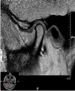
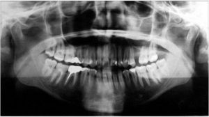
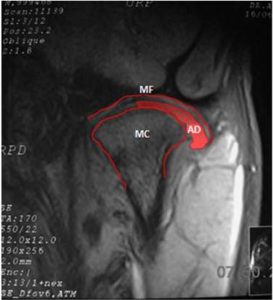
Comparative studies between MRI and OPG In order to study the disorders diagnosed by the MRI, and its correlation with the vertical mandibular asymmetries (VMA) viewed in the OPG, 200 images of magnetic resonance imaging in oblique planar coronal sections and of orthopantomogram of people, both in maxillomandibular occlusion position taken with the same equipment and in the same place were analyzed and evaluated. The ESECi database analyzed belongs to patients who attended to the center between 2004 and 2007, and who contributed with the necessary information for the analysis. This research conceals not only people’s identity but also any data that could be used to identify them, according to Habeas Data in force in Argentina. (Figures 1 and 2).  In the MRI images, the following anatomical structures: mandibular condyle (MC), articular disc (AD) and mandibular fossa (MF) need to be distinguished to later recognize the joint alterations. The medial and/or lateral disc displacement (MDD and/or LDD) in relation to the MC was specifically identified.

507487-fig-1

Figure1: MRI of Right TMJ in Oblique Sagittal Planar Section


507487-fig-2

Figure 2: Orthopantomogram

 
Traces were drawn on the images of MRI oblique coronal sections of TMJ to evaluate the TMD, and on the images of OPG to recognize the asymmetry. The methodology used in the MRI and the OPG was the following:

Tracing methods on MRI: 1) The MC, the MF and the AD are located. 2) The MC and MF perimeters, and the AD outline are traced.

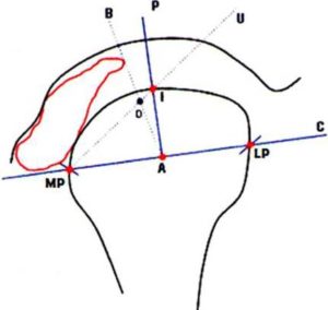
MRI Planning: 1) the most convex point in the medial pole (MP) and lateral pole (LP) of the MC is drawn. If one or both poles are flat, the intermediate point of the surface is marked. 2) A line joining the MP and LP points is traced creating a plane C. 3). On plane C, point A is equidistantly marked between the MP and LP points. 4) A perpendicular line to plane C from point A (Plane P) intersecting the cortical of the mandibular condyle is traced creating point I. 5) A line (U) from the MP point (with a medial articular disc displacement) or from point LP (with a lateral articular disc displacement) to point I is traced. 6) A line (B) from point A to the border of the medial or lateral articular disc is traced according to its displacement creating an intersection point D with plane U.

There is a disc displacement when point D is equidistant from the distance I-MP/LP, or farther in a medial or lateral way (according to the displacement). (Figures 3 and 4).

507487-fig-3

Figure 3: MRI in Oblique Planar Coronal Section on which the Following Structures are Recognized: Mandibular Fossa (MF), Mandibular Condyle (MC) and Articular Disc (AD)

507487-fig-4

Figure 4: Planes on Traces in MRI to Establish the Medial-Lateral Displacement of the Articular Disc

 
Vertical mandibular asymmetries diagnosis: to avoid inclinations provoked by the cranial position in the orthopantomograph and the diverse 3D craniofacial dimensions, which are very difficult to control, — Panmekiate et al. (1995), López López et al — the measurements are registered in millimeters, but expressed in percentages to establish the differences between vertical mandibular symmetry or asymmetry in the OPG.

The millimeter dimensional measurement in the OPG is done on the traces measuring the vertical height of the ramus establishing vertical asymmetry when the bilateral difference is higher than 3%. Panmekiate et al., López López et al.

Tracing methods on OPG: 1) the mandibular bone perimeter is located. 2) A line is traced following the bone cortical through the inferior border of the mandibular body (basal bone) from the mental symphysis to the right and left, mandibular angle and posterior border of the ramus to the neck of the condyle delimiting the posterior, superior and anterior borders of the condyle. The line continues in the border of the mandibular notch to end up tracing the posterior border, and the vertex of the coronoid process.

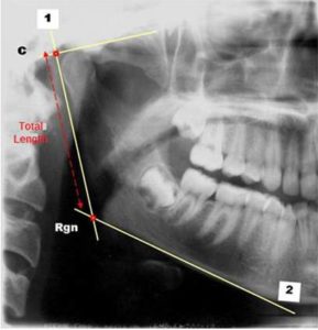
OPG Planning: 1) A tangent is traced on the posterior border of the ramus (plane 1). — 2) A tangent is traced on the inferior border of the mandibular body (plane 2). — 3) Point C is determined as a result of the intersection between plane 1 and a perpendicular line traced on the superior point of the MC. — 4) The retrognation point (Rgn) is identified in the intersection of planes 1 and 2.

The total length of the ramus is measured from point C to Rgn point. (Figure 5).

 
507487-fig-5

Figure 5: Traces on OPG to Measure the Ramus Length

 
The magnitudes of the vertical mandibular asymmetries and the articular disc displacement are established in percentages.

Results

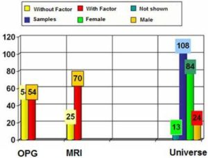
108 cases (84 females and 24 males) were studied. In 13 cases it was not possible to observe the AD of the TMJ not being part of the correlation between the VMA and the functional TMD. (Figure 6).

507487-fig-6

Figure 6: Column Chart: Represents the Total and the Distribution of the Studied Cases. – Without Factor: Control Cases; With Factor: Problem Cases; Not Shown: Cases in which the AD is Not Seen; Samples: Total Number of Studied Cases in the Research; OPG: Orthopantomogram; MRI: Magnetic Resonance Imaging

 
A random result of 54 cases with vertical radiographical asymmetry (OPG- A) and 54 cases with normal or vertical radiographical symmetry (OPG- N) were obtained. To carry out the statistical analysis, the value 0 (zero) was assigned to the vertical radiographical symmetry of the ramus and value 1 (one) to the asymmetry. 70 of these people had temporomandibular internal functional disorders (TMD) and 25 had normal temporomandibular joints (NTMJ) valued 1 and 0 respectively in the statistic correlation. The cases with factor (problem cases) were represented by the TMJ with TMD (medial or lateral articular disc displacement) and the vertical mandibular asymmetries (OPG-A). The cases without factor (control cases) were represented by the normal temporomandibular joint (NTMJ) and the vertical mandibular symmetry (OPG-N).

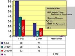
When the TMD and the VMA were correlated, 70 TMJ presented medial-lateral disc displacement or functional TMD (1-MRI), 38 of which had a vertical mandibular radiographical asymmetry (OPG-1) and 32 had symmetry (OPG-0). 10 out of 25 TMJ without functional TMD (0-MRI) had vertical mandibular asymmetries (OPG-1) and 15 had symmetries (OPG-0). (Figure 7).

507487-fig-7

Figure 7: Column Chart. Crossing of Variables of Control and Problem Cases. Result of the Kendall’s W Test Statistical Analysis

 
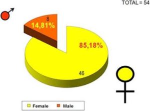
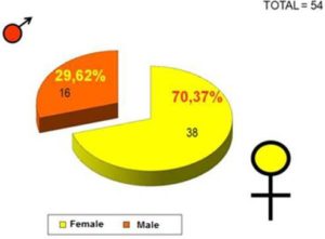
Once this data was analyzed with Kendall’s W Test statistic value — coefficient of concordance 0,206, degree of freedom 1 and P  ≤ 005, the obtained value was 0,0001 indicating  a non-significant statistic agreement between the functional TMD and the VMA. Symmetry/asymmetry distribution according to gender: 46 females and 8 males with radiographical mandibular symmetry (Figure 8), and 38 females and 16 males with radiographical mandibular asymmetry. (Figure 9).

507487-fig-8

Figure 8: Correlation between Gender and Vertical Mandibular Symmetry

 
507487-fig-9

Figure 9: Correlation between Gender and Vertical Mandibular Asymmetry

Discussion

The current research proves the existence of TMD by MRI and shows that 40% of people have TMD and vertical mandibular asymmetry. However, Kopp et al. (1979) reports a larger asymmetry of the vertical condylar height measured by panoramic radiograph in patients with clinical TMD; Habets et al. (1988) informs a statistically significant difference between patients with routine teeth groups, and clinically diagnosed patients under treatment of craniomandibular disorders on the vertical condylar asymmetry. Like Bezuur et al (1988, 1989) established, Kopp et al (1979) and Habets et al (1988) based on the TMD clinical diagnosis, pointed out that 74% of the patients with TMD have more than 3% of vertical condylar asymmetry. The differences found in this research could arise from the method used to diagnose the TMD. The authors quoted above carried out a clinical diagnosis, while this study was based on MRIs.

Standard panoramic radiographs could not be accurate when compared to the OPG of cone beam computed tomography (CBCT). Sunny Young H. (2005) and Rezende Barbosa GL, Nascimento MD, Ladeira DB, Bomtorim VV, da Cruz AD, Almeida SM. (2013) have observed a significant dimensional difference with higher precision in the CBCT. According to the results of this research, the OPG is not an option when compared to the CBCT since it does not provide the exact internal joint condition.

When Winocur E, Reiter S, Krichmer M, Kaffe I (2010) compare the OPG with the MRI to prove the existence of a degenerative joint disease, the radiographic technique was accurate in only a 20%. As proved in our research, the OPG diagnosis based on the vertical mandibular asymmetry was not relevant.

Conclusions
 
The significant differences found in this study between a functional TMD and the asymmetry shown by the OPG support the current recommendations, that panoramic radiographs should not be a routine exam to evaluate TMD, but it is the first option when there is a potential dental problem.

Subsidy

Ministry of Science and Technology Res. N: 1187/2007 (Government of the province of Córdoba — Argentina) Tutoring Programme for Research Teams.

Acknowledgment

Ministry of Science and Technology of the Government of the province of Córdoba, Argentine Republic.

References
 

Anantharam, B., Chahal, N., Stephens, N. & Senior, R. (2010). “Temporo-mandibular Joint Dislocation: An Unusual Complication of Transoesophageal Echocardiography,” European Journal of Echocardiography, 11 (2), 190-1.
PublisherGoogle Scholar

Bezuur, J. N., Habets, L. L. M. H. & Hansson, T. L. (1988). “The Recognition of Craniomandibular Disorders- A Comparison between Clinical, Tomographical, and Dental Panoramic Radiographical Findings in Thirty- One Subjects,”Journal of Oral Rehabilitation, 15 (6), 549-54.
PublisherGoogle Scholar

Bezuur, J. N., Habets, L. L. M. H. & Hansson, T. L. (1989). “The Recognition of Craniomandibular Disorders; Condylar Symmetry in Relation to Myogenous and Arthrogenous Origin or Pain,” Journal of Oral Rehabilitation, 16 (3), 257-60.
PublisherGoogle Scholar

Giambartolomei, L. A. (2003). “Anatomía del Complejo Articular Cráneomandibular — ATM,” Ed: Publicaciones Universidad Nacional de Córdoba; ISBN: 950 — 33 — 0410 — 5.
Publisher

Habets, L. L. M. H., Bezuur, J. N., Naeiji, M. & Hansson, T. L. (1988). “The Orthopantomogram, an Aid in Diagnosis of Temporomandibular Joint Problems. ll. The Vertical Symmetry,” Journal of Oral Rehabilitation, 15 (5), 465-71.
PublisherGoogle Scholar

Hutchinson, S. Y. (2005). “Cone Beam Computed Tomography Panoramic Images vs. Traditional Panoramic Radiographs,” American Journal of Orthodontics & Dentofacial Orthopedics, 128 (4), 550-550.
PublisherGoogle Scholar

Isberg, A. (2003). ‘Disfunción de la Articulación Temporomandibular,’ Ed. Artes Médicas Latinoamérica, 91, 173 – 180.
Google Scholar

Kopp, S. & Rockler, B. (1979). “Relationship between Clinical and Radiographic Findings in Patients with Mandibular Pain or Dysfunction,” Acta Radiologica: Diagnosis, 20, 456.
PublisherGoogle Scholar

Larheim, T. A. (2005). “Role of Magnetic Resonance Imaging in the Clinical Diagnosis of the Temporomandibular Joint,”Cells Tissues Organs 180 (1), 6-21.
PublisherGoogle Scholar

López López, J., Chimenos Küstner, E., Blanco Carrión, A., Reselló Llabrés, X. & Jané Salas, E. (2005). “Diagnóstico Por la Imagen de los Trastornos de la Articulación Craneomandibular,” Avances en Odontoestomatología, 21 (2), 71-88.
PublisherGoogle Scholar

Okeson, J. P. (1999). ‘Anatomía Funcional y Biomecánica del Sistema Masticatorio. En: Tratamiento de Oclusión y Afecciones Temporomandibulares,’ Ed. Mosby,  7-17.

Paesani, D.,  Westesson, P.- L., Hatala, M. P., Tallents, R. H.  & Broods, S. L. (1992). “Accuracy of Clinical Diagnosis for TMJ Internal Derangement and Arthrosis,” Oral Surgery, Oral Medicine, Oral Pathology, 73 (3), 360-3.
PublisherGoogle Scholar

Panmekiate, S., Petersson. A., Rohlin, M., Akerman, S. & Isberg, P.- E. (1995). “Corrected Sagittal Tomography of the Temporomandibular Joint. Influence of Errors in Film and Patient Positioning on Linear and Angular Measurements,” Acta Odontologica Scandinavica 53 (4), 264-9.
PublisherGoogle Scholar

Park, J. W., Song, H. H., Roh, H. S., Kim, Y. K. & Lee, J. Y. (2012). “Correlation between Clinical Diagnosis Based on RDC/TMD and MRI Findings of TMJ Internal Derangement,” International Journal of Oral & Maxillofacial Surgery 41(1), 103-8.
PublisherGoogle Scholar

Pasquet, G. & Cavezian, R. (2009). “Diagnostic Means Using oral and Maxillofacial Cone Beam Computed Tomography: Results,” Journal de Radiologie 90 (5 Pt 2), 618-23.
PublisherGoogle Scholar

Reyez Tellez Girón, J. & Nuñez Tovar, C. (1998). ‘Nomenclatura Anatómica Internacional,’ Ed. Médica Panamericana, 19, 59, 44-45.
Google Scholar

Rezende Barbosa, G. L., Nascimento, M. D. C. C. &  Ladeira, D. B. S., Bomtorim, V. V., da Cruz, A. D. & Almeida, S. M. (2013). “Accuracy of Digital Panoramic Radiography in the Diagnosis of Temporal Bone Pneumatization: A Study in Vivo Using Cone-Beam-Computed Tomography,” Journal of Cranio-Maxillofacial Surgery. Jul 11. pii: S1010-5182(13)00178-9.
PublisherGoogle Scholar

Winocur, E., Reiter, S., Krichmer, M. & Kaffe, I. (2010). “Classifying Degenerative Joint Disease by the RDC/TMD and by Panoramic Imaging: A Retrospective Analysis,” Journal of Oral Rehabilitation, 37(3):171-7. 
PublisherGoogle Scholar

Shares