Fracture Resistance in Fiber Reinforced Composite Restorations — An in Vitro Study

Flavia Călburean1, Bogdan Mihai Gălbinașu2, Roxana Cara-Ilici2 and Ion Pătrașcu3

1Orthodontics, “Carol Davila” University of Medicine and Pharmacy, Bucharest, Romania

2Prosthesis Technology and Dental Materials, “Carol Davila” University of Medicine and Pharmacy, Bucharest, Romania

3”Carol Davila” University of Medicine and Pharmacy, Bucharest, Romania

Academic Editor: Fernanda De Carvalho Panzeri Pires-de-Souza

Cite this Article as:

Flavia Călburean, Bogdan Mihai Gălbinașu, Roxana Cara-Ilici and Ion Pătrașcu (2014), "Fracture Resistance in Fiber Reinforced Composite Restorations — An in Vitro Study," JMED Research, Vol. 2014 (2014), Article ID 669406, DOI: 10.5171/2014.669406

Copyright © 2014 Flavia Călburean, Bogdan Mihai Gălbinașu, Roxana Cara-Ilici and Ion Pătrașcu. Distributed under Creative Commons CC-BY 3.0

Abstract

The objective of our study was to expand knowledge on the versatility of dental composites as they make a reliable armamentarium for the clinician, whether he is restoring small dental defects or large cavities at the limits of conservative treatment. For this paper we have tested In vitro a possibility to enhance fracture resistance in lateral composite restorations by reinforcing them with glass fibers; as it was hypothesized that a glass fiber mesh would increase the cohesion of a composite restoration, thus its fracture resistance.  Extracted sound teeth received a large composite restorations reinforced with a fiber glass net, and then subjected to vertical load until colapse. The results were that reinforced fillings resisted more to vertical load than conventional fillings and the remaining dental tissues suffered less from the failure of the restoration. Glass fiber reinforcement enhances intrinsic resistance of composite restorations and the restored tooth as a whole.
Keywords: Composite restorations, fracture resistance, fiber glass reinforcement.

Introduction

Composite dental materials have revolutionized restorative dentistry due to their conservative technique, adhesive bond to natural tissues and their adequacy for aesthetic refurbishment [17, 18, 19, 20].

Massive lateral restorations, as a daily routine, do not cease to raise issues in terms of technique and predictability [6, 8, 14]. There are many incontrolable factors that influence the longevity of these restorations: occlusal forces, malfunction, chemically active food and drinks, temperature and humidity variations, salivary or bacterial enzymes etc. Some materials [4, 5, 9] are specially fabricated to meet lateral restoration needs, but their efficiency is not always as expected. Thus, the clinician often confronts himself with a difficult treatment decision between a more or a less conservative method in order to comply with other therapeutical principles: prophylactic, biomechanical, ergonomic and financial also.

Seldom there is a distinct indication for a large direct composite filling or an inlay or a crown. Whatever the decision, the clinician takes some risk. One may choose to restore a large cavity in a fast and cheap way with a direct composite filling; however, the tooth is exposed to fracture along with the restoration failure. On the other hand, a crown would be more expensive and time consuming, and although it is credited with longevity, it may as well endanger the tooth considering stealth loosening of the crown, root cavities and restricted endodontic access etc [18].

For this paper we have looked upon the possibility of enhancing fracture resistance in lateral composite fillings for endodontically sane teeth. We have studied, in vitro, how the incorporation of parodontal use glass fibers change the mechanical behavior of these restorations.

It was hypothesed that reinforcing restorations would increase their resistance by absorbing the forces occurring inside the material subjected to stress. Furthermore, a layer of fiberglass placed at the interface filling – deep cavity wall could stop / redirect line fractures occured in the composite and propagated up to this level, deviating the path of fractures that could engage remaining dental tissues — the most serious consequence of direct composite restorations failure.

It is known that glass fibers are resistant to tension and are able to stop the propagation of fractures in the composite mass [10]. Theoretically, cracks initiated in the restoration are stopped or deflected by glass fibers, and are no longer transmitted to the cervical region. In addition, the distribution of stress within the fiber reinforced restoration may increase the strength of the restoration. The distribution of the fibers may be unidirectional or bi-directional thereby providing resistance in one or two space dimensions.

To the date, research in this field [3, 10, 16, 23, 24] has brought suggestive results for the opportunity of reinforcement, yet not conclusive. Recently, conducted studies cover only little of reinforcement techniques uses, and have delt with endodonticaly treated samples only.

For our study, we have hypothesed that glass fiber reinforcement of the restorations would increses their load resistance by increasing the cohesion of the composite material.

If the use of fiberglass products for composite filling reinforcement proves reliable, this technique could enrich the therapeutic arsenal of restorative dentistry. The next step would be to make available fiber glass products specifically designed for this purpose.

Materials and Methods

A total of 27 extracted sound teeth (15 premolars and 12 molars) were stored for a period of 2-4 months in distilled water, at room temperature, in the dark, after prior disinfection with chloramine solution (0. 5% for 10 days).

Teeth were divided into three equal groups who underwent the following treatments: the first batch (A) were fully preserved, the second group (B) were drilled resulting in large cavities which have been filled with dimetacrilic resin composite. In the third group (C) large fillings have been reinforced by applying a layer of glass fiber mesh disposed on the pulp wall of the cavity. For groups B and C, large cavities were drilled with respect to the pulp chamber, which included the palatal cusp in premolars, the disto-oral in molars with four cusps, respectively one vestibular cusp in three cusp molars. A simulated cusp fracture complication was added to the MOD cavity. VO size of the cavities was a third of the intercusp distance, measured with the callipers (fig. 1).

669406-fig-1

Fig. 1: Preparation for a 3 Cusp Molar

 
Cervical wall was placed at 1 mm from the cement-enamel junction and measured 1 mm in depth. The depth of the cavities was established according to the average tooth substance above the pulp chamber ceiling [1, 2], keeping a 1.5 mm layer of dentin to it.

For all preparations, we used a rounded tip diamond burr, under continuous cooling water from the turbine.

The teeth in group B were classically filled with composite. We chose an adhesive system that requires total removal of remaining dentinal debris. We differentially etched enamel (30 s) and dentin (15 s) with 37% phosphoric acid semi-gel (Bisco).

After thorough washing and drying, we used primer and bonding adhesive system PQ1 Bonding Agent (Ultradent) applied for 20 s. After air thinning, the adhesive was cured for 30 s. We then applied packable composite A 3, 5 shaded Premise (Kerr Corporation) in oblique layers of 2 mm, in contact with as few cavity walls as possible. Each layer was cured for 40s. We rebuilt the side walls and the occlusal aspect with respect for morphological criteria.

Teeth in group C were subjected to demineralization, priming and bonding maneuvers, and then we applied a thin layer of flowable composite Premise Flowable (Kerr Corporation) and a piece of fiberglass mesh PFM 3 (Dentapreg) 3mm x 2mm of 0, 5 mm thickness (Fig. 2).

669406-fig-2
Fig. 2: Fiber Glass Net Placement
 
The product contains bidirectional interleaved fibers (at an angle of 120°), silanised and resin impregnated. Flowable composite and impregnated glass fibers were simultaneously light-cured. Next, we applied composite filling in 2 mm layers until the correct morphology of the tooth was achieved (Fig. 3).

669406-fig-3
Fig. 3: The Final Restoration in a Molar
 
During the next phase of the study, all 3 groups were simultaneously subjected to a thermo cycling regimen in LTC unit 100 (fig. no. 4). This device consists of two baths in which water can be maintained at a constant temperature, a container device that moves the samples from one bath to the other, passing through a drying stage. We have carried out 500 cycles according to the following thermo cycling protocol [22]:

  • 20 seconds immersion in 550 C bath
  • 10 seconds drying
  • 20 seconds immersion in 50 C bath


669406-fig-4

Fig. 4: The Termocycling Device

 
After thermo cycling, the samples were prepared for the testing phase: the teeth roots were embedded in acrylic resin (Duracryl, Spofadental) using a cylindrical mold. For holding the samples during the experiment, we used a metal shank made of two halves forming a cylindrical cavity with adaptable diameter when put side by side (Fig. 5).

669406-fig-5

Fig. 5: Molar Ready to Receive Vertical Loading

 
All samples were vertically loaded by means of a conical piece [7] fixed to the device Testomatic metal (Benchmarking, England). Testomatic (Fig. 6) is a modern testing apparatus which provides parameter adjustment by means of a computer and also digital recording and interpretation of the test results in the form of variation graphs.

669406-fig-6
Fig. 6: Tooth Subjected to Occlusal Loading
 
Thus, the machine was set to exert a constant pressure at a speed of 1 mm / min, and to stop when the resistance of the sample decreases sharply by 25%, which corresponded to the fracture of the sample. Each sample was then labeled and stored individually together with the disconnected fragments.

Each specimen was subjected to a macrovisual analysis based on the following: the extent of tooth fracture (pulp chamber or enamel-cement junction engagement), type of fracture (adhesive or cohesive fracture of the fillings) and fracture line trajectory (i.e. its location as respects to dental tissues).

669406-fig-7
Fig. 7: Cracked Sound Tooth
 
 
The analysis consisted of macroscopic assessment of trajectory lines of fracture (Fig. 7). Conventionally, a glossy and heterogeneous fracture surface similar to primed and bonded dental tissues was considered an adhesive fracture. Similarly, a heterogeneous fracture surface, but of matte finish, coupled with a matching surface on a detached fragment was interpreted as an invasive tooth fracture. A smooth homogenuous fracture surface A 3, 5 shaded was interpreted as being the result of cohesive fracture in the filling.

All the phases of this study were documented with photographs, and software for thermo cycling and compressive loading tests could provide roadmaps of operations (Fig. 8).

669406-fig-8
Fig. 8: Variation of Load Resistance
 
 
Results

For control group A, the compression tests lead to the following results:


  • Premolars have resisted 981, 5 N  mean (sd 196)
  • Molars opposed a resistance of 2360, 4 N  mean (sd 924)
  • Most sound premolars (three out of four) experienced fractures that did not involve the root or the pulp chamber. In molars, 3 teeth experienced fractures localized on the cusps and two molars suffered vertical crown-root fractures.


For group B (classical composite fillings) the results were:


  • Premolars have resisted 615, 75 N mean (sd 242.5)
  • Molars opposed a resistance of 1240 N  mean (sd 374)
  • Premolar fractures resulted equally from adhesive and cohesive failures. In none of the cases did the fracture line exceed the composite restoration-dental tissue interface.
  • Molars generally showed fracture lines continued in the dental tissues (Fig. 9) causing outstanding cusp fractures (three cases), fractures below the cement-enamel junction (two cases) or fractures opening the pulp chamber (one case).
  •  
669406-fig-9
Fig. 9: Fracture Line Continued in the Dental Tissues
 
Teeth in group C (composite restorations reinforced with fiberglass mesh) had the following results:

  • Mean compressive strength of 946 N in premolars (sd 536), with 53, 63% more than premolars in group B
  • Mean compressive strength of 1595, 4 N for molars (sd 306), with 28, 33%  more than molars in group B
  • Most fractures (14 walls out of 22) were due to the adhesive failure of the restorations, and in no circumstance did the fracture line interested remaining dental hard tissues (Fig. 10).  
669406-fig-10
Fig. 10: Failure of Reinforced Fillings
 
669406-tab-1,2

Extensive classical restorations in premolars resisted to a relatively small load, 615. 75 N on the average, while the reinforced restorations exhibited more fracture strength (946 N), but these results did not reach the statistical significance level (p= 0, 1889 student’s T-test).

Most fractures walls in teeth with reinforced fillings were adhesive to a greater extent than in conventional fillings, which suggests an increase in the internal resistance of fillings, i. e. greater cohesion, in addition to a strength boost of the assembly (p=0, 019 Fisher test).

Molars with composite fillings have proved a medium strength of 1240 N, and in molars with fiber glass reinforced fillings compressive strength was 1599 N. The influence of fiber reinforcement on these data seems obvious but still it does not have statistical relevance (p=0, 2418 Student’s T-test).

In 3 of the 5 molars classically restored with composite resins, the fracture of the filling spread in the remaining natural tissues (cusps, tissues beyond the CEJ), while in molars with reinforced restorations, the remaining tooth structure suffered no damage. The association of fiber reinforcement and lack of tooth structure injury proved to some extent to be significant statistically (p=0, 083 Fisher test).

Generally, the adhesive fractures occurred in the pulp wall underneath the fiber mesh. So, there was no disjunction between the glass fibers and the fluid resin applied above it. This result is suggestive of the reliability of the connection between silanised glass fibers and composite resins. However, the fracture line separated fragments of glass fiber mesh, but this is probably due to the fact that the fibers were woven in a loose manner.

Discussion

The present study received attention for standardization, proper technique and the use of approved devices employment [7, 21, 22]. There were, however, empirical stages (e.g. macroscopic analysis of fracture surfaces) or non-quantifiable aspects (i.e. the contact surface between the specimen and the metal cone piece that was exerting the load). Also, the flowable composite used for accommodation of the glass fiber in reinforced restorations may have contributed to the better resistance to load of these samples [15]. The high values for standard deviation in group A and B in molars, and in group C in premolars are due to one aberrant data in each of these that would have been ignored in a larger set of data.

In this study we have used endodontically sound teeth with an original preparation inspired from the authors’ practices.

This restorative technique with the use of glass fibers does not require significant time, and the cost increases. The handling and application of glass fibers do not require increased manual skills; however, the doctor may need an assistant.

Practical experience in this study has made the authors aware of some features that a reinforcing material designed specifically for direct composite resin restorations should have:

  • Fibers should be woven in a mesh, thin enough to be pliable and adaptable to the shape of the cavity floor
  • Fiber beams should be thin but tightly woven to form 90 ° angles
  • The product should be small for a single application, packaged in opaque containers
  • The kit should contain a transparent instrument with which fibers are maintained during the polymerization by allowing curing light to pass through.


Glass fiber reinforcement of direct restorations proves to be a promising solution for large crown restorations, as it enhances intrinsic resistance of composite restorations and the restored tooth as a whole. The way in which the application of a layer of glass fibers produce these improvements suggest the need for finite element analysis, in order to observe any changes in the concentration of stress compared to what is known in the conventional composite restorations.

Conclusion

While entirely aware of the limits of our study, we can conclude that restored teeth have significantly less resistance to veritical load than sound teeth (e. g. 2360.4 N average sound molars and 1246N average restored molars), but fiber reinforced restorations resist at greater forces than classical fillings (28, 33% more in molars and 53, 63% more in premolars). Also, fiber reinforced restorations have greater cohesion, and they fail mostly by adhesion colapse (p=0, 019), which proves less harmful for the remaining natural tissues (0, 083).

References

1. Deutsch, A. S. & Musikant, B. L. (2004). “Morphological Measurements of Anatomic Landmarks in Human Maxillary and Mandibular Molar Pulp Chambers,” Journal of Endodontics, 30 (6) 388-390.
PublisherGoogle Scholar

2. Deutsch, A. S., Musikant, B. L., Gu, S. & Isidro, M. (2005). “Morphological Measurements of Anatomic Landmarks in Pulp Chambers of Human Maxillary Furcated Bicuspids,” Journal of Endodontics 31 (8) 570-573.
PublisherGoogle Scholar

3. Belli, S., Cobankara, F. K., Eraslan, O., Eskitascioglu, G. & Karbhari, V. (2006). “The Effect of Fiber Insertion on Fracture Resistance of Endodontically Treated Molars with MOD Cavity and Reattached Fractured Lingual Cusps,”Journal of Biomedical Materials Research 79 (1) 35-41.
PublisherGoogle Scholar

4. Bowen, R. L. & Rodriguez, M. S. (1962). “Tensile Strength and Modulus of Elasticity of Tooth Structure and Several Restorative Materials,” Journal of American Dental Associacion, 64 378-387.
PublisherGoogle Scholar

5. Braga, R. R., Hilton, T. J. & Ferracane, J. L. (2003). “Contraction Stress of Flowable Composite Materials and Their Efficacy as Stress-Relieving Layers,” Journal of American Dental Associacion 134 721—728.
PublisherGoogle Scholar

6. Brunthaler, A., Lucas, T., Sperr, W. & Schedle, A. (2002). ‘Longevity of Direct Resin Composite Restorations in Posterior Teeth,’ Received Published online: 27 May 2003.

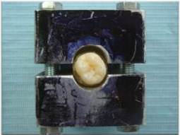
7. Ciocan, L.T., Iordache, B., Gălbinașu, B. & Pătrașcu, I. (2012). Invention Brevet no 122801 ‘Equipment for Testing the Cohesion of Mixed Dental Materials,’ State Office of Invention Nd Trademark, România.

8. Çötert, H. S., Sen, B. H. & Balkan, M. (2001). “In Vitro Comparison of Cuspal Fracture Resistances of Posterior Teeth Restored with Various Adhesive Restorations,” International Journal of Prosthodontics 14 374—378.
PublisherGoogle Scholar

9. Craig, G. R. (2001). ‘Materiale Dentare Restauratoare,’ All Educațional, București, România.

10. Fennis, W. M. M., Kreulen, C. M., Tezvergil, A., Lassila, L. V. J., Vallittu, P. K. & Creugers, N. H. J. (2011). “In Vitro Repair of Fractured Fiber-Reinforced Cusp-Replacing Composite Restorations,” International Journal of Dentistry, article ID 165938.
PublisherGoogle Scholar

11. Fusuyama, T. (1980). ‘New Concepts in Operative Dentistry: Differentiating Two Layers of Carious Dentin and Using an Adhesive Resin,’ Quintessence Publishing Co, Chicago, Il.
Google Scholar

12. Gatin, E., Matei, E., Pîrvu, D. A., Gălbinașu, B. M. & Iordache, S. (2012). “Comparative Survey of the Most Used Self Adhesive Dental Cements Based on Resin Composites,” Digest Journal of Nanomaterials and Biostructures, 7 (1) 207-214.
PublisherGoogle Scholar

13. Gălbinașu, B. M., Miculescu, F., Pîrvu, D., Ciocan, L. T., Herschbach, D., Tonko, A., Pătrașcu, I. & Stoiciu, F. (2012). ‘Dental Tissue — Inlay Interface Structural Shift at Mechanical and Thermal Stresses,’ Optoelectronics and Advanced Materials 6 (3-4) 459-464.
Google Scholar

14. Hickel, R. & Manhart, J. (2001). “Longevity of Restorations in Posterior Teeth and Reasons for Failure,” The Journal of Adhesive Dentistry 3 (1) 45-64.
PublisherGoogle Scholar

15. Cara, R. R., Fleming, G. J. P., Palin, W. M., Walmsley, A. D. & Burke, F. J. T. (2007). “Cuspal Deflection and Microleakage in Premolar Teeth Restored with Resin-Based Composites with and without an Intermediary Flowable Layer,” Journal of Dentistry 35(6) 482-4894.
PublisherGoogle Scholar

16. Kuijs, R. H., Fennis, W. M. M., Kreulen, C. M., Roeters, F. J. M., Creugers, N. H. J. & Burgersdijk, R. C. W. (2006). “A Randomized Clinical Trial of Cusp-Replacing Resin Composite Restorations: Efficiency and Short-Term Effectiveness,” International Journal of Prosthodontics 19(4):349-54 PubMed PMID: 16900817.
PublisherGoogle Scholar

17. Malhotra, N., Mala, M. &  Acharya, S. (2001). “Resin-Based Composite as a Direct Esthetic Restorative Material,”Compendium of Continuing Education in Dentistry 32(5) 14-24.
PublisherGoogle Scholar

18. Margeas, R. (2012). “Composite Resin: A Versatile, Multi-Purpose Restorative Material,” Compendium of Continuing Education in Dentistry 33(1) 25-27.
PublisherGoogle Scholar

19. Ritter, A. V. (2001). “Posterior Resin-Based Composite Restorations – Clinical Recommendations for Optimal Success,” Journal of Esthetic and Restorative Dentistry, 13 88—99.
PublisherGoogle Scholar

20. Ritter, A. V. (2008). “Posterior Composites Revisited,” Journal of Esthetic and Restorative Dentistry 20 (1) 57-67 PubMed PMID: 18237342.
PublisherGoogle Scholar

21. Sarrett, D. C. (2005). “Clinical Challenges and the Relevance of Materials Testing for Posterior Composite Restorations,” Dental Materials 21(1) 9-20.
PublisherGoogle Scholar

22. Technical Specifications ISO/T6 1405:2003 (E) ‘Dental Materials- Testing of Adhesion to Tooth Structure,’

23. Tezvergil, A., Fennis W. M. M., Kuijsc, R. H., Lassilab, L. V. J., Kreulen, C. M., Creugersa, N. H. J. & Vallittub P. K. (2004). “In Vitro Fracture Resistance of Fiber Reinforced Cusp-Replacing Composite Restorations,” Dental Materials 21 (5) 565-572.

24. Xu, H. H. K. et al. (2003). “Continuous-Fiber Preform Reinforcement of Dental Resin Composite Restorations,” Dental Materials, 19 (6) 523-530.
PublisherGoogle Scholar

Shares