Introduction
As a free radical, nitric oxide (NO) reacts with O2- to produce OH- and NO2- , which are more toxic than NO and contribute to disturbing the function of the respiratory chain, induction of DNA damage and cell apoptosis and aggravation of ischemia-reperfusion injury (Ito et al.,2010). Under physiological condition, there is no expression of inducible nitric oxide synthase (iNOS). The release of local excitatory amino acid (EAA) was increased after cerebral ischemia-reperfusion, which activates iNOS and induces NO synthesis (Choi et al., 2010; Wang et al., 2011). This effect is associated with lipid peroxide (Caso et al., 2008; Bi et al., 2009) and induction of cell apoptosis or death (Brown et al.,2010, Perluigi et al., 2010; Rey et al., 2011). After cerebral ischemia, increased production of free radical promoted degeneration and necrosis of vascular endothelial cell, aggravated infarction edema and destroyed reconstruction of blood supply in infarct area (Bailey et al., 2004). Under normal circumstances, the presence of effective antioxidant system is required to maintain the physiological levels of free radicals. Glutathione peroxidase (GSHPx) can convert H2O2 into H2O and eliminate the toxic action of oxygen free radical (Yan et al., 2008; Wang et al., 2010). Cell culture assay shows that picroside II can reduce damage of H2O2-induced PC12 cells and improve cell survival rate (Li et al.,2002, Tao et al., 2003; Guo et al., 2007). Picroside II, which is an iridoid glucoside, is an active constituent extracted from the traditional Chinese medicine (TCM) hu-huang-lian, Animal experiments indicate that picroside II might regulate the expressions of iNOS, SOD and Caspase-9 (Li et al., 2010; Sun et al., 2011) to inhibit the neuronal apoptosis induced by cerebral ischemia reperfusion injury and improve the neurological function of animals (Li et al., 2010; Li et al., 2010). Author’s former research indicated that the optimized therapeutic dose and time window is injecting picroside II intraperitonenally with 10-20mg/kg body weight at ischemia 1.5h in cerebral ischemic injury in rats (Pei et al., 2012). The authors aimed to further explore the optimum therapeutic dose and time window according to the quantitative measurement of NO and GSHPx level in blood and brain homogenates.
Materials and Methods
Animal Models
A total of 78 adult healthy male Wistar rats, SPF grade, weight 230—250 g, were supplied by the Experiment Animal Center of Qingdao Drug Inspection Institute (SCXK (LU) 20100010). All animals were given time to adapt to the laboratory environment, allowed free access to food and water in a housing with natural illumination for a week. The room temperature was controlled at 23 ± 2°C. All rats were randomly divided into sham-operation group with 10 rats, model group with 10 rats and treatment group with 48 rats. All animals were fasted 12h before operation. The rats were anesthetized by injecting intraperitoneally 10% chloral hydrate (300 ml/kg) and fixed in supine position to conduct aseptic operation. Global forebrain ischemia models were established by bilateral common carotid artery occlusion (BCCAO) of rats (Márquez-Martín et al., 2012). The successful rat models of global forebrain ischemia were internalized into the experiment group (ten rats that unconsciousness or dead 2 hours after operation were excluded). The rats in the sham operation group underwent the same surgical procedures except BCCAO operation. The Local Ethical Committee of Experimental Animals of Medical College of Qingdao University approved that all the steps of this study were compliant with ethical rules.
Orthogonal Experimental Design
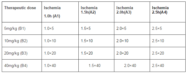
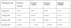
Total 48 successful global forebrain ischemia rat models of treatment group were internalized into the experiment and divided randomly according to the principle of orthogonal experimental design of [L16(45)] consisting of two impact factors with four impact levels (Table 1). The impact factor A is the therapeutic time widow designed four levels: 1.0 h, 1.5 h, 2.0 h, 2.5 h after ischemia. The impact factor B is the therapeutic drug dose which has four levels as follows: 5 mg/kg, 10 mg/kg, 20 mg/kg and 40 mg/kg body weight. The orthogonal experiment was repeated 3 times.
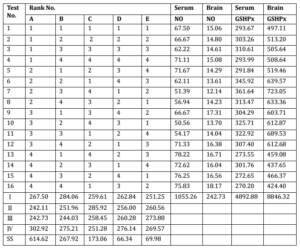
Table 1: Orthogonal Experimental Design of [L16 (45)]
Treatment Methods
Picroside II (Tianjin Kuiqing Medical Technology Co., Ltd., CAS No: 39012-20-9, purity >98%, Molecular formula: C23H28O13, Molecular weight: 512.48) was diluted into 1% solution with saline solution and injected intraperitoneally according to the corresponding designed doses and ischemia time (Pei et al., 2012) in the orthogonal layout [L16(45)]. Rats in sham group were simultaneously suffered the same doses saline solution.
Sample Collection
After treatment with picroside II for 24h, total of rats were anesthetized with 10% chloral hydrate (300 ml/kg) andcollected 4ml blood through heart to centrifuge at 4000 rpm for 10 min to separate serum, and stored at −20℃. And then the rats were perfused with 200ml saline solution through heart. The brain was taken out completely through fast craniotomy and the olfactory bulb and prefrontal brain tissue were excised. Five hundreds milligram brain tissue were prepared backward from optic chiasma (Bregma 0.00mm), grinded to power in pre-cooling mortar and then homogenized by ultrasonic wave to extract cellular proteins with lysis buffer (500ul lysis buffer + 5ul PMSF, No.P0013, Beyotime Institute of Biotechnology. Shanghai, China) according to the proportion of 1:3, centrifuged with refrigerated centrifuge (Model Eppendorf 5801, Germany) at 12,000 r/min for 10 min at 4℃. The supernatant liquid was collected and stored at −20℃. The protein concentration in the supernatant was determined by the BCA method (Wuhan Boster Biological Engineering Co., Ltd., China).
Detection Index
The NO levels and GSHPx activities of serum and brain homogenate were measured by method of nitrate reductase and chemical colorimetry. According to the kit (Nanjing Jiancheng biology technology company) instructions, the serum or brain tissue homogenate were resolved at room temperature and centrifuged. The 100μl supernatant liquid was collected. We measured absorbance value with UV spectrophotometer (Beckmann DU640, USA) at a wavelength of 550nm (NO) and 412nm (GSHPx) and then calculated the content of NO (μmol/L) content and the activity of GSHPx (U/ml).
Statistical Analysis
SPSS 17.0 software was used for data statistical analysis. According to the result, multi-group comparison was made by analysis of orthogonal test whether different level of administrating time dose and the therapeutic time window.
Results
Measurement Results
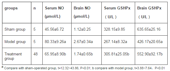
In model group, the contents of NO in serum and brain tissue homogenate were increased significantly than those in sham-operated group and the activities of GSHPx were obviously decreased than those in sham-operated group(t=12.32~43.86, P<0.01). In treatment group, the contents of NO in serum and brain tissue homogenate were obviously decreased than those in model group and the activities of GSHPx were increased significantly than those in model group(t=3.88~7.64, P<0.01. See tables 2 and 3.
Table 2: The Measurement Results Results of NO and GSHPx
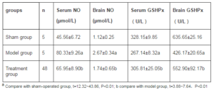
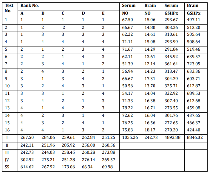
Table 3: L16 (45) Orthogonal Layout and the Results of Measurement
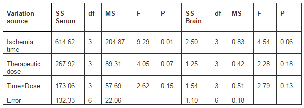
ANOVA Analysis of NO Content
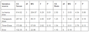
There was significant difference (P0.05) found among various therapeutic dose (factor B) and various interaction between medication time and therapeutic dose (factor C). Pairwise comparisons among data of all independent groups by least-significant-difference (LSD) method showed that there was significant difference (P0.05). There was significant difference of NO content between therapeutic dose 5mg/kg (B1) and 20mg/kg (B3) (P 0.05). Therefore, according to the principle of lowest therapeutic dose with longest time window, A2B3 is the best combination group, i.e., the optimized time window and therapeutic dose is ischemia 1.5h with 20mg/kg body weight.
There was no significant difference (P > 0.05) of NO content in brain tissue among various medication time (factor A), various therapeutic dose (factor B) and various interaction between medication time and therapeutic dose (factor C). Pairwise comparisons among data of all independent groups by least-significant-difference (LSD) method showed that there was significant difference (P<0.05) of NO content between medication time 1.0h (A1) and 1.5 h (A2), 1.5 h (A2) and 2.5 h (A4), while there was no significant difference between each other for the rest groups ((P > 0.05). There was no significant difference (P>0.05) among various therapeutic dose. Therefore, according to the principle of lowest therapeutic dose with longest time window, A2B3 is the best combination group, i.e., the optimized time window and therapeutic dose is ischemia 1.5h with 20mg/kg body weight.
Table 4: ANOVA Analysis of NO Content
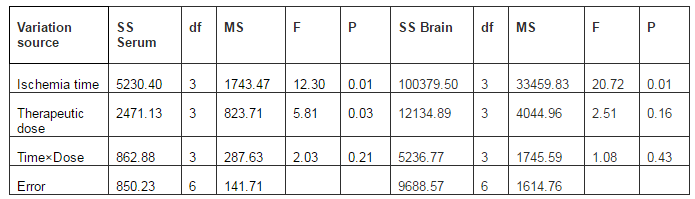
ANOVA Analysis of GSHPx Activity
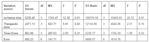
There was significant difference (P0.05). Pairwise comparisons among data of all independent groups by least-significant-difference (LSD) method showed that there was no significant difference (P>0.05) of the activity of GSHPx in serum between medication time (ischemia time) 1.0h (A1) and 2 h (A3), 1.5 h (A2) and 2 h (A3), but significant difference existed between each other for the rest groups (P 0.05), but a significant difference existed between each other for the rest groups (P < 0.05). Therefore, according to the principle of lowest therapeutic dose with longest time window, A2B2 is the best combination group, i.e., the optimized time window and therapeutic dose is ischemia 1.5h with 10mg/kg body weight.
There was significant difference (P0.05). Pairwise comparisons among data of all independent groups by least-significant-difference (LSD) method showed that no significant difference ( P > 0.05) of the activity of GSHPx in brain tissue between medication time 1.0h (A1) and 2.5 h (A3), 1.5 h (A2) and 2.0 h (A3), while there was significant difference between each other for the rest groups (P < 0.05). There was significant difference of the activity of GSHPx in brain tissue between therapeutic dose 5mg/kg (B1) and 20mg/kg (B3) (P 0.05). Therefore, according to the principle of lowest therapeutic dose with longest time window, A3B3 is the best combination group, i.e., the optimized time window and therapeutic dose is ischemia 2.0h with 20mg/kg body weight.
Table 5: ANOVA Analysis of GSHPx Activity
Discussion
Modern medical studies show that the active ingredients of traditional Chinese medicine have good neuroprotective effect. Salvia miltiorrhiza (Zhang et al., 2010) and calycosin (Guo et al., 2012) could reduce mitochondrial injury induced by cerebral ischemia-reperfusion in rats, significantly decrease contents of MDA (Malondialdehyde) and protein hydroxyl compounds, upregulate the activity of SOD (Superoxide dismutase), CAT (Catalase) and GSHPx, and at the same time, inhibit the expression of 4-Hydroxy-2-nonenal. Chrysophanol formulations (Li and Zhang et al., 2010) could significantly increase the activities of GSHPx in whole blood and SOD in plasma in rats with cerebral ischemia reperfusion, and improve the prognosis in ischemic cerebrovascular disease. Liquiritin (Sun et al., 2010) could decrease the content of MDA in brain tissue of mice and increase the activities of CAT and GSHPx. Salidroside (Yan et al., 2008) can obviously reduce the content of NO in rats with cerebral ischemia reperfusion, and improve the damage of neurons induced by ischemia reperfusion, the mechanism of which may be the enhanced expression of downstream step in pathways of intracellular mitogen-activated protein kinase (Li et al., 2002; Li et al., 2000). Cell culture assay shows that picroside II has obvious protective effect on damage of H2O2-induced PC12 cells, which has a relationship with effect of direct scavenging oxygen free radicals and enhancing cell itself function of antioxidant system (Tao et al., 2003). Experiments on animal models indicate that picroside II could improve the antioxidant capacity of brain tissue in cerebral ischemic injury in rats, reduce the oxidative damage induced by cerebral ischemia-reperfusion and improve nerve behavior function of rats (Zhao et al., 2010).
As the detection index of this experiment, NO can directly reflect changes of free radical content in cerebral ischemic injury and GSHPx can reflect the change of antioxidant content in cerebral ischemic injury. In this experiment, the authors designed four time points at 1 h, 1.5 h, 2 h and 2.5 h after brain ischemic injury in rats, and inject intraperitoneally picroside II with four therapeutic doses of 5 mg/kg, 10 mg/kg, 20 mg/kg and 40 mg/kg. The experiment was carried out according to orthogonal table of [L16(45)] to explore the best therapeutic dose and best time window of picroside II in treating cerebral ischemic injury by determining the expressions of NO and GSHPx. The results indicate that there is significant difference in therapeutic effect among various medication times and various therapeutic doses of picroside II. But the results are not consistent with the best combination of different indexes.
Conclusion
According to the principle of lowest therapeutic dose with longest time window, the optimized therapeutic dose and time window should be injecting picroside II intraperitonenally with 10-20mg/kg body weight at ischemia 1.5-2.0h.
Acknowledgments
This work is supported by National Natural Science Foundation of China (Grant No. 81041092) and Natural Science Foundation of Shandong Province (Grant No. ZR2011HM050).
References
Bailey, D. M., Kleger, G. R., Holzgraefe, M., Ballmer. P. E. & Bärtsch, P. (2004). “Pathophysiological Significance of Peroxidative Stress, Neuronal Damage and Membrane Permeability in Acute Mountain Sickness,” Journal of Applide Physiology, 96(4)1459-1463.
Publisher – Google Scholar
Bi, X., Yan, B., Fang, S., et al. (2009). “Quetiapine Regulates Neurogenesis in Ischemic Mice by Inhibiting NF-kB p65/p50 Expression,” Neurological Research, 31(2)159-166.
Publisher – Google Scholar
Brown, G. C. (2010). “Nitric Oxide and Neuronal Death,” Nitric Oxide, 23(3)153-165.
Publisher – Google Scholar
Caso, J. R., Pradillo, J. M., Hurtado, O., Leza, J. C., Moro, M. A. & Lizasoain, I. (2008). “Toll-Like Receptor 4 Is Involved in Subacute Stress-Induced Neuroinflammation and in the Worsening of Experimental Stroke,” Stroke,39(4) 1314-1320.
Publisher – Google Scholar
Choi, D. W. (1988). “Glutamate Neurotoxicity and Disease of the Nervous System,” Neuron, 1(8) 623-634.
Publisher
Guo, C., Tong, L., Xi, M., Yang, H., Dong, H. & Wen, A. (2012). “Neuroprotective Effect of Calycosin on Cerebral Ischemia and Reperfusion Injury in Rats,” Journal of Ethnopharmacology,144(3)768-774.
Publisher – Google Scholar
Guo, M. C., Cao, Y. & Liu, J. W. (2007). ‘Protective Effects of Picroside â…¡ on Glutamate Injury of PC12 Cells,’ Chinense Journal of Clinical Pharmacology and Therapeutics,12(4) 440-443.
Ito, Y., Ohkubo, T., Asano, Y., Hattori, K., Shimazu, T.,Yamazato, M., Nagoya, H., Kato, Y. & Araki, N. (2010). “Nitric Oxide Production during Cerebral Ischemia and Reperfusion in eNOS and nNOS-Knockout Mice,” Current Neurovascular Research, 7(1) 23-31.
Publisher – Google Scholar
Li, C., Zhang, D. S., Zhao, X. Q., Shi, F. Y., Zhu, C. L. & Xue, G. P. (2010). “Experimental Screening Study of Three Chrysophanol Formulations on Learning and Memory Function of Mice with Cerebral Ischemia Reperfusion,” Chinese Pharmacological Bulletin, 26(12)1607-1612.
Publisher – Google Scholar
Li, P., Matsunaga, K. & Ohizumi, Y. (2000). “Nerve Growth Factor Potentiating Compounds from Picrorhizae Rhizome,”Biological and Pharmaceutical Bulletin, 23(7)890-892.
Publisher – Google Scholar
Li, P., Matsunaga, K., Yamakuni, T. & Ohizumi, Y. (2002). “Picrosidesâ… and â…¡, Selective Enhancers of the Mitogen-Activated Protein Kinase-Dependent Signaling Pathway in the Action of Neuritogenic Substances on PC12D Cells,” Life Sciences, 71(15) 1821-1835.
Publisher – Google Scholar
Li, Q., Guo, Y. L., Li, Z. & Xu, X. Y. (2010). “The Interference of Picroside II on the Expressions of Caspase-3 and PARP Following Cerebral Ischemia Reperfusion Injury in Rats,” Chinese Pharmacological Bulletin, 26(3) 342-345.
Publisher – Google Scholar
Li, Q., Li, Z., Xu, X. Y., Guo, Y. L. & Du, F. (2010). “Neuroprotective Properties of Picroside II in Rat Model of Focal Cerebral Ischemia,” International Journal of Molecular Sciences, 11(11) 4580-4590.
Publisher – Google Scholar
Li, Z., Li, Q., Guo, Y. L., Qin, L. H. & Luan, L. J. (2010). ‘Interference Effect of Picroside II on Cerebral Ischemia Reperfusion Injury in Rats,’ Acta Anatomica Sinica, 41(1) 9-12.
Márquez-Martín, A., Jiménez-Altayó, F., Dantas, A. P., Caracuel, L., Planas, A. M. & Vilak, E. (2012). “Middle Cerebral Artery Alterations in a Rat Chronic Hypoperfusion Model,” Journal of Applied Physiology, 112(3) 511-518.
Publisher – Google Scholar
Pei, H. T., Su, X., Zhao, L., Li, H. Y., Guo, Y. L., Zhang, M. Z. & Xin, H. (2012). “Primary Study for the Therapeutic Dose and Time Window of Picroside II in Treating Cerebral Ischemic Injury in Rats,” International Journal of Molecular Sciences, 13(2) 2551-2562.
Publisher – Google Scholar
Perluigi, M., Di Domenico, F., Giorgi, A., Schininà , M. E., Coccia, R., Cini, C., Bellia, F., Cambria, M. T., Cornelius, C., Butterfield, D. A. & Calabrese, V. (2010). “Redox Proteomics in Aging Rat Brain: Involvement of Mitochondrial Reduced Glutathione Status and Mitochondrial Protein Oxidation in the Aging Process,” Journal of Neuroscience Research, 88(16) 3498-3507.
Publisher – Google Scholar
Rey, B., Roussel, D., Teulier, L., Eyenga, P., Degletagne, C., Belouze, M. & Duchamp, C. (2011). “Functional Argument for the Existence of an Avian Nitric Oxide Synthase in Muscle Mltochondria:Effect of Cold Acclimation,” FEBS Lett,585(1)173-177.
Publisher – Google Scholar
Sun, L., Li, X. D., Wang, L., Qin, L. H., Guo, Y. L. & Zhou, Z. (2011). ‘The Anti-Oxidant Effect and the Possible Mechanism of Picroside â…¡ in Cerebral Ischemia Reperfusion Injury in Rats,’ Neural Regeneration Research, 6(15) 1141-1146.
Sun,Y. X., Tang,Y., Wu, A. L., Liu, T., Dai, X. L., Zheng, Q. S. & Wang, Z. B. (2010). “Neuroprotective Effect of Liquiritin against Focal Cerebral Ischemia/Reperfusion in Mice via Its Antioxidant and Antiapoptosis Properties,” Journal of Asian Natural Products Research, 2(12)1051-1060.
Publisher – Google Scholar
Tao, Y. W., Liu, J. W., Wei, D. Z., Su, W. & Zhou, W. Y. (2003). ‘Protective Effect of Picroside- II on the Damage of Cultured PC12 Cells in Vitro,’ Chinense Journal of Clinical Pharmacology and Therapeutics, 8(1) 27-30.
Google Scholar
Wang, J. Y., Yu, R. & Yao, M. H. (2010). “Protective Effects and Mechanisms of Salidroside on Cerebral Ischemia-Reperfusion Injury,” China Journal of Traditional Chinese Medicine and Pharmacy, 25 (3) 456-459.
Publisher – Google Scholar
Wang, L.- Q., Jia, J., Guo, M.- F., Zhang, Y., Zhang, X.- Y., Guo, J-. J., Geng, R.- J., Li, B., Zhong, H. & Li, D.- L. (2011). “Effect of L-Arginine Amino Guanidine and Agmatine on Focal Cerebral Ischemia-Reperfusion Injury in Rats,”Chinese Journal of Pathophysiology, 27(6) 1213-1217.
Publisher
Yan, T. H., Jia, Y., Yang, W. & Wang, Q. J. (2008). ‘Protective Effects of on Focal Cerebral Ischemic Reperfusion Injury in Rats,’ Chinese Pharmacological Bulletin, 24(11) 1521-1524.
Zhang, Q. L., Sun, Y. B., Wang, H. Y., Song, S. J. & Bai, B. (2010). “Effects of Salvia Miltiorrhiza Bunge. F. Alba. on Mitochondrial Damage and Apoptosis Induced by Cerebral Ischemia and Reperfusion,” Athophysiology, 26(4)725-729.
Publisher – Google Scholar
Zhao, D. M., Zhang, Z. Q., Duan, Y. X., Zhang, B. Z. & Liu, Q. S. (2010). ‘Neuroprotective Effect of Picroside II on Cerebral Ischemia-Reperfusion Injury in Rats,’ Journal of International Pharmaceutical Research, 37(6)461-468.
Google Scholar