Introduction
Impacted tooth is the tooth that does not appear in the oral cavity on expected time and typically there is no possibility for growth for a variety of reasons, such as high density of bone over tooth, growth inhibition by adjacent tooth, mucosal high thickness on growing tooth or genetic causes that stopped the normal growing. (Al-Khateeb et al. 2006, Hupp et al. 2008)
Mandibular third molar is the most common impacted tooth, and the surgery for its removing by oral and maxillofacial surgeons is the most common performed surgical procedure (Obeichina et al. 2001) and might be due to pathological changes or prophylactic purposes (Polat et al. 2008) .With regard to complications of impacted teeth such as periodontal problems, dental caries of the adjacent tooth, root resorption of adjacent teeth, crowding, cyst or tumor formation and pain with unknown cause, a well planed, accurate and in time treatment should prevent next complications (Aitasalo et al. 1972).
Various studies are done about impacted third molar and related complications. A study by Obiechina et al. assessed the symptoms and the patterns of mandibular third molar impaction in Nigeria. They observed 338 patients between ages of 16 and 54 (Mean 24.4±6.1 years old). They had 473 mandibular impacted third molars (Obeichina et al. 2001). In another study by Aitasalo et al., the panoramic radiography of 4063 patients in the Institute of Dentistry of Turku University was studied. Impacted teeth were found in %14.1 patients. The third molar tooth had the highest impaction prevalence (76.1%) (Aitasalo et al. 1972). In another similar study in 1000 patients, Quek et al. studied the prevalence of impacted third molars in 68.6% of radiographs (Quek et al. 2003). As jaws in different populations vary in size and this affects the impaction of the third molars, this study was done for the evaluation of the presence of the impacted third molars, and their complications in the panoramic radiography of patients in a group of Iranian population over 20 years old who referred to oral and maxillofacial radiology of Babol dental faculty (North of Iran).
Methods and materials
In this cross-sectional study, 2000 panoramic radiographs from archives of oral & maxillofacial radiology, department of Babol university of medical sciences (north of Iran), related to patients older than 20 years old, were observed by an oral & maxillofacial radiologist. Digital radiographs were stored in computer. Pictures were taken from conventional radiographs on negatoscop by a digital photography camera (Olympus, Japan, 12Mpixels), and then were stored in the computer. Images were evaluated on a computer screen. The radiographs of the patients with impacted teeth were selected and information about age and gender of patients, and presence of impacted teeth were recorded in an information form, and characteristics of impacted teeth were examined. All teeth that were not erupted for any reasons in the oral cavity, were evaluated and considered as impacted teeth.
Characteristics and methods of evaluation of each radiograph were as following:
* The patient’s gender is defined as male or female.
* Age of patients who are older than 20 years.
* Number of impacted third molars which could be from 1 to 4 teeth.
* Position of impacted third molar in comparison to the second molar angle longitudinal axis can be mesial, distal, vertical, horizontal or reversed.
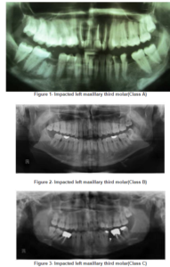
* Impacted depth, in comparison to the occlusal surface of the adjacent second molar, can be placed in one of the positions A, B or C; Class A (The occlusal plane of the impacted tooth is at the same level as the adjacent tooth); Class B (The occlusal plane of the impacted tooth is between the occlusal plane and the cervical line of the adjacent tooth); Class C (The occlusal plane of the impacted tooth is apical to the cervical line of the adjacent tooth) (Hupp et al. 2008 , Aitasalo et al. 1972).
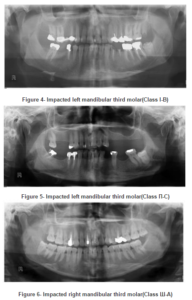
* Relation of the mandibular impacted third molar to the anterior border of ramus, according to the amount of mesiodistal width of the impacted tooth that is covered by ramus, will be placed in one of class I or II or III. Class I 3rd molar impaction: Situated anterior to the anterior border of the ramus. Class II 3rd molar impaction: Crown ½ covered by the anterior border of the ramus. Class III 3rd molar impaction: Crown fully covered by the anterior border of the ramus. (Hupp et al. 2008 ,Obeichina et al. 2001)
* Root development of impacted third molars, expressed as if either they are complete or incomplete.
* Defects (cavities) of distal margin of the second adjacent molar will be expressed as presence or absence.
* The presence or absence of alveolar bone resorption
* Relationship between roots of lower third molar and mandibular canal expressed in the form of direct or indirect relationship. If any of the following cases are happening, the relationship is considered to be directly:
1. Absence of lamina dura on places where the canal is associated with the third molar
2. Radiolucent band width roots of the third molar
3. Thinning of mandibular canal where the third molar root passes
4. Any change of the canal on the location of third molar
5. Third molar root deviation by canal. (Howe et al. 1960)
After completion of the questionnaires, data were analyzed by SPSS18 software and Mann-Whitney test. P<0.05 was significant.
Results
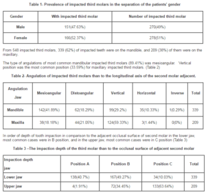
In the present study, 333 (16.65%) of the patients had impacted tooth. 313 of these patients (15.65%) had just impacted wisdom tooth, 16 patients (8.0%) just had other impacted tooth, and 4 patients (2.0%) had impacted wisdom teeth and other impacted teeth. A total of 317 (85.15%) cases of the 548 patients had impacted wisdom tooth. The frequency of patients with impacted wisdom tooth and the number of impacted wisdom tooth in both genders are given in Table 1.


(Figures 1,2,3). In order of relation of the impacted third molar to anterior border of ramus, it was concluded that 183(53.98%) impacted third molars were in class I, 146(43.06%) were in class II and 10 (2.96%) teeth were in class III. (Figures 4, 5, 6)
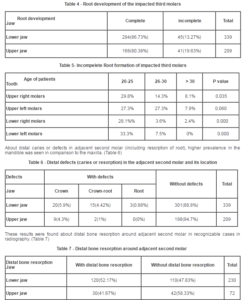
In order of root development of the impacted third molars, most of them had complete root. Incomplete root formation was showed in table 5. (Table 4, 5)

The relationship between the root of the lower impacted third molar with the inferior alveolar canal from 287 cases recognizable in radiography, 109 (37.98%) of them had direct relationships, and 178 (62.02%) of them had indirect relationships with the inferior alveolar canal .
Among 333 patients who had an impacted tooth, 20 patients had impacted teeth other than impacted third molar. They had 23 impacted teeth (9 canine teeth at the upper jaw , 3 canine teeth at the lower jaw , 3 second premolar teeth in the lower jaw , 1 second molar tooth in the lower jaw (figure 1 ), and 7 added impacted teeth were at premolars region).
Discussion
In the present study, 16.65% of patients had impacted teeth, and most impacted tooth was the third molar. In the study of Aitasalo et al., panoramic radiography of 4063 patients was evaluated. Impacted teeth in 14.1% of patients were found and the most common impacted tooth was the third molar (76.1%). There were no differences in maxilla and mandible regarding prevalence. Also they found no differences in prevalence of impacted third molars according to gender (Aitasalo et al. 1972). Also in our study, the frequency of impacted third molars was not equal on the maxilla (38%) and mandible (62%), which is similar to findings in the results of studies of Saglam (Saglam et al. 2003), and Chu (Chu et al. 2003). Also, the frequency of impacted third molars was not equal in women (52.37%) and men (47.63%), which can be due to more space deficiency in the posterior of mandible and smaller size jaws in women. Also, it can be due to racial differences and differences of jaws’ size between patients of two studies. However, more studies are needed in this area (north of Iran) to obtain more accurate results.
The results of this study showed that the prevalence of impacted third molar teeth in women was higher than that of men, which is compatible with the results of Haghanifar’s study. (Haghanifar et al. 2006) In the study of Quek et al., prevalence of impacted teeth was reported in women more than in men, too (Quek et al. 2003).
In the study of Obiechina, the evaluation of impaction status using Pell & Gregory classification showed that 358 (54.55%) impacted teeth were in position A, 151(31.92%) teeth were in position B, and 64 (13.53%) cases were in position C. 107(22.62%) cases were in class I, 288 (60.89%) in class II, and 78 (16.49%) cases were in class III (Obiechina et al. 2001). These results are different compared to those of our study in that the mandible, position of impaction in most of the cases was in position B (49.27%) and class I (53.98%), which can be due to differences of mandible size in the populations of both studies or racial differences. Also, it can be due to the age of patients that in our study it was over 20 years old but in the study of Obiechina it was over 16 years old. (Obiechina et al. 2001) These findings were different from the findings of Byahatti (Byahatti et al. 2012) and Obiechina’s (Obiechina et al. 2001), and were similar to the study of Sandhu (Sandhu et al. 2005).
In the study of Azaz on 200 mandibular impacted third molars, 60% of them had distinct relations to inferior alveolar canal nerve, 19% had true relations and 41% were as superimposition. The majority of the molars showed complete root formation in the third decade of life (Azaz Purcha et al. 1976).
In the present study, according to relations between mandibular impacted third molar and the canal in radiographically detectable cases, 109 (37.98%) cases had contact to canal and 178 (62.02%) cases did not have any contact. This can be due to differences in explanation methods of relation to canal, because we considered the teeth with superimposition on canal, assumed as indirect relationship. In the study of Azaz (Azaz Purcha et al. 1976) regarding the radiographic evaluation that accompanies the surgical procedure, we can say that their study was more accurate.
In the study of Gupta et al, most of the 988 mandibular impacted third molars had upright position (39.93%). Most of the impacted third molars were in position A (61.84%) and class II (79.65%). Also, there was true relation between mandibular canal and root of impacted third molar in 211(21.35%) cases (Gupta et al. 2011). We found that most of impacted third molars had mesioangular position (41.89%) that is different from the results of Gupta’s study. It can be due to space deficiency in the distal of second molar for correct positioning of the third molar. Also, angulation and depth of impaction of impacted third molar are important in the difficulty of surgery.
In the study of Sheikh, the prevalence of distal caries of the second molar adjacent to the impacted third molar was evaluated. Out of 200 evaluated impacted third molars, there was distal caries of the second molar in 42.5% of cases. Fifty one percent of the third molars had mesioangular impaction (Sheikh et al. 2012). In this study, the prevalence of distal caries of the second molar (11.2% in mandible and 5.3% in maxilla) was much less than their study, which can be due to differences in the hygienic level of both studied populations. Also, according to these findings, it seems that the angulation of impaction is important in distal caries of second molar, because in our study the angulation of impacted teeth in the mandible was mesioangular in most cases (41.89%) and, compared to maxilla whose angulations of third molars were upright in most cases (59.33%), the prevalence of distal caries of second molars were more prevalent.
According to the resorption of the distal alveolar bone of the adjacent second molar, in radiographically detectable cases, 52.17% of mandibular cases had bone resorption and 47.83% of the cases did not have it. Also in the maxilla, 41.67% of cases had bone resorption and 58.33% did not have it. Also, we cannot detect the status of second molar distal bone by using the two dimensional radiography in all the cases.
Conclusion
In the present study, 16.65% of patients had impacted teeth. The prevalence of impacted third molars in women was more than men which can be due to differences in the size of their jaws. But, this needs more studies to get more accurate results. So, we should take more care about evaluating impacted third molars and making decision about their prophylactic removal in women. The prevalence of impacted third molars in the mandible was higher than that of the maxilla, which can be due to more space deficiency in the posterior mandible compared to maxilla, but this needs more studies and evaluations to obtain more accurate results.
Most of mandibular third molars had mesioangular position, and in the maxilla most of the cases were upright. So, we can say that in the mandible, the possibility of eruption and positional correction of impacted wisdom teeth is much less than maxilla and this mostly shows the importance of impaction in the mandible.
Finally, we can say that regarding the high percentage of the resorption of the distal bone of the second molar and their distal caries (defects) that have clinical importance, we can suggest the prophylactic removal of impacted wisdom teeth.
Acknowledgment
Acknowledgment
The present study is the result of a research project No. 8930341, approved by the Research Council of Babol University of Medical Sciences, and the thesis of a dentistry student — Dr. Rashid Soufizadeh. The authors would like to thank the Deputy of Research and Technology of Babol University of Medical Sciences for financially supporting this project.
References
1. Aitasalo, K. Lehtinen, R. and Oksala, E. (1972) “ An orthopantomographic study of prevalence of impacted teeth”, Int J Oral Surg 1(3) 117-20 .
Publisher – Google Scholar
2. Al-Khateeb, T.H. and Bataineh, A.B. (2006) “Pathology associated with impacted mandibular third molars in a group of Jordanians”, J Oral Maxillofac Surg, 64 (11) 1598-602.
Publisher – Google Scholar
3. Azaz purcha, B. Shteyer, A. and Piamenta, M. (1976) “Radiographic and clinical manifestations of the impacted mandibular third molar” Int J Oral Surg 5(4) 153-160.
Publisher – Google Scholar
4. Byahatti, S. and Ingafou, M. (2012) “Prevalence of eruption status of third molars in Libyan students”, Dent Res J (Isfahan) 9(2) 152-7.
Publisher – Google Scholar
5. Chu, F.C. Li, T.K. Lui, V.K. Newsome, P.R. Chow, R.L. Cheung, L.K. (2003) “Prevalence of impacted teeth and associated pathologies — a radiographic study of the Hong Kong Chinese population”, Hong Kong Med J 9 (3): 158-63.
Google Scholar
6. Gupta, S. Bhowate, R.R. Nigam, N. and Saxena, S. (2011) “Evaluation of impacted mandibular third molars by panoramic radiography”, ISRN Dent 2011, 406714.
7. Haghanifar, S. and Emamverdizadeh, P.(2006) “Radiographic evaluation of impacted teeth prevalence Dental faculty of Babol 2004-2006”, Journal of Ghasr-e-Baran 1(1) 14-17.
8. Hupp, J.R. Ellis Ш, E. and Tucker, M.R. (2008) Contemporary oral and maxillofacial surgery, Mosby Elsevier, St Louis, Missouri, USA.
9. Howe, G.L. and Poyton, H.G. (1960) “Prevention of damage to the inferior dental nerve during the extraction of mandibular third molars”, Br Dent J 109 355-63.
10. Obiechina, A.E. Arotiba, J.T. and Fasola, A.O. (2001) “Third Molar Impaction: evaluation of the symptoms and pattern of impaction of mandibular third molar teeth in Nigerians”, OdontoStomat Tro, 24 (93) 22-5.
11. Polat, H.B. Ozan, F. Kara, I. Ozdemir, H. and Ay, S. (2008) “Prevalence of commonly found pathoses associated with mandibular impacted third molars based on panoramic radiographs in Turkish population”, Oral Surg Oral Med Oral Pathol Oral Radiol Endod 105 (6) e41-7.
Publisher – Google Scholar
12. Quek, S.L. Tay, C.K. Tay, K.H. Toh, S.L. Lim, K.C. (2003) “Pattern of third molar impaction in a Singapore Chinese population: a retrospective radiographic survey”, Int J Oral Maxillofac Surg 32 (5) 548-52.
Publisher – Google Scholar
13. Sağlam, A.A. and Tüzüm, M.S. (2003) “Clinical and radiologic investigation of the incidence, complications and suitable removal times for fully impacted teeth in the Turkish population”, Quintessence 34 (1) 53-9.
Google Scholar
14. Sandhu, S. and Kaur, T. (2005) “Radiographic evaluation of the status of third molars in the Asian-Indian students”, J Oral Maxillofac Surg 63(5) 640-5.
Publisher – Google Scholar
15. Sheikh, M.A. Riaz, M. and Shafiq, S. (2012) “Incidence of distal caries in mandibular second molars due to impacted third molars–A clinical and radiographic study” Pakistan Oral & Dental Journal 32(3) 364-370.


