Proximal Tibiofibular Synostosis, an Unexpected Complication of a Proximal Tibial Osteotomy

I. Esenkaya and K. Unay 

Department of Orthopaedic and Traumatology, Istanbul Medeniyet University Goztepe Research and Training Hospital, Istanbul, Turkey

Academic Editor: Michel P. J. van den Bekerom

Cite this Article as:

I. Esenkaya and K. Unay (2013), "Proximal Tibiofibular Synostosis, an Unexpected Complication of a Proximal Tibial Osteotomy," International Journal of Case Reports in Medicine, Vol. 2013 (2013), Article ID 113410,
DOI: 10.5171/2013.113410

Copyright © 2013 I. Esenkaya and K. Unay. Distributed under Creative Commons CC-BY 3.0

Abstract

Proximal tibiofibular synostosis is rare. A proximal tibiofibular synostosis occurred because too long implant screw passed through the lateral cortex in a patient who had undergone a proximal open wedge osteotomy for right knee medial compartment osteoarthritis. The patient had no complaints about synostosis after implant removal. No proximal tibiofibular synostosis due to proximal tibial medial open wedge osteotomy has been reported. We report a new cause of proximal tibiofibular synostosis and a new complication of proximal tibial osteotomy.  
Keywords: Synostosis; tibia; fibula; osteotomy; complication.

Introduction

One treatment for medial knee osteoarthritis is a proximal tibial osteotomy, which can involve a closed wedge, open wedge or dome osteotomy. These surgical techniques give rise to various complications, including osteonecrosis of the proximal fragment, fracture of the proximal fragment, medial or lateral cortical fracture, intra-articular fractures extending to the medial or lateral plateau, nonunion, delayed union, popliteal artery injury, peroneal nerve injury, wound healing problems, haematoma, incomplete correction, slope changes, deformity relapse, collateral ligament injuries, stiff knee, implant failure, pain due to the implant, deep venous thrombosis, pulmonary embolism, arterial thrombosis, compartment syndrome, septic arthritis and infection [1-12].

Proximal tibiofibular synostosis is rare. Synostosis at this site may be congenital [13], idiopathic [14], secondary to osteochondroma [15], secondary to multiple hereditary exostosis [16] or caused by trauma [17]. To our knowledge, no proximal tibiofibular synostosis due to proximal tibial medial open wedge osteotomy has been reported. We report a new cause of proximal tibiofibular synostosis and a new complication of proximal tibial osteotomy.

Case Report

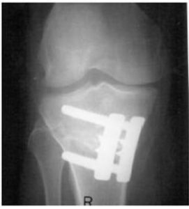
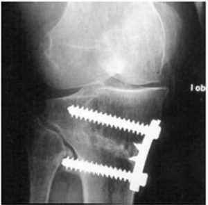
A proximal tibial open wedge osteotomy was performed on a 62-year-old woman with right knee medial compartment osteoarthritis (Fig 1). A longutidal incision from lower pole of patella to tuberositas tibia was used for surgery. Special plates were used designed by the first author [2, 3]. During the surgical procedure, drill penetrated the lateral cortex. No additional complication was seen in the intraoperative and early postoperative periods. Weight bearing allowed at 2.5 month. At the 10-month follow-up, the beginning of a proximal tibiofibular synostosis due to the distal anterior screw of the osteotomy plate was seen (Fig 2). Since the patient had no pain, screw removal was refused.

113410-fig-1

Figure 1: Early Postoperative Anteroposterior X-Ray

113410-fig-2
Figure 2: Anteroposterior X-Ray 10 Months Postoperatively

 
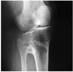
A follow-up X-ray 13 months postoperatively showed more of the screw in the internal oblique view than in the true anteroposterior view and the synostosis was evident (Fig 3). The condition was explained to the patient, who was convinced to undergo implant removal surgery. The implant was removed, with no surgical intervention done on the synostosis.

113410-fig-3

Figure 3: Internal Oblique X-Ray 13 Months Postoperatively

 
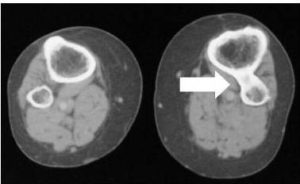
The patient has been followed for 61 months after implant removal surgery. During follow-up, no pain occurred due to medial compartment osteoarthritis or proximal tibiofibular synostosis (Fig 4 and 5). Inform consent was obtained from the patient.

113410-fig-4

Figure 4: Internal Oblique X-Ray after Implant Removal


113410-fig-5

Figure 5: Bilateral Proximal Tibiofibular Computed Tomography; the White Arrow Shows the Right Knee Synostosis

 
Discussion

An asymptomatic proximal tibiofibular synostosis secondary to the long distal anterior screw in a proximal tibial osteotomy plate was reported.

The reported symptoms of a symptomatic proximal tibiofibular synostosis include pain at the proximal tibiofibular junction [18], pain in the ipsilateral ankle [14], restricted range of motion in the ipsilateral ankle [13], angular deformity [19] and leg length discrepancy [20]. Our patient was asymptomatic, unlike reported cases, which was why we did not perform any surgical intervention for the synostosis.

A proximal tibial osteotomy is an important treatment alternative for medial knee compartment osteoarthritis. Like any surgical technique, this technique has some complications. To our knowledge, no tibiofibular synostosis has been reported in the English literature after a proximal tibial osteotomy.

Follow-up X-rays are important for diagnosing such complications. Anatomically, the proximal tibia is shaped like a triangular prism, and an internal oblique X-ray is necessary to determine the length of the implant screws in the lateral proximal cortex after a medial open wedge proximal tibial osteotomy. This is taken with the X-ray beam positioned anteroposteriorly and the lower extremity in 30—40° of internal rotation. This position gives a true lateral view of medial tibial implants (Fig 3).

Tibiofibular synostosis due to an implant has been reported in the distal tibiofibular joint [21-23]. However, we could find no report of a proximal tibiofibular synostosis due to an implant screw in the English literature.
    
In conclusion, proximal tibiofibular synostosis may occur when a long implant screw is used in a proximal tibial open wedge osteotomy. The internal oblique X-ray is important for the diagnosis. During follow-up, no surgical intervention may be required.

References

1. Aglietti, P., Buzzi, R., Vena, L. M., Baldini, A. & Mondaini, A. (2003). “High Tibial Valgus Osteotomy for Medial Gonarthrosis: A 10- to 21-Year Study,” The Journal of Knee Surgery. 16:21-6
PublisherGoogle Scholar

2. Esenkaya, I. & Elmali, N. (2006). “Proximal Tibia Medial Open-Wedge Osteotomy Using Plates with Wedges: Early Results in 58 Cases,” Knee Surgery, Sports Traumatology, Arthroscopy. 14:955-61.
PublisherGoogle Scholar

3. Esenkaya, I. (2005). “Fixation of Proximal Tibia Medial Opening Wedge Osteotomy Using Plates with Wedges,” Acta Orthopaedica et Traumatologica Turcica. 39:211-23
PublisherGoogle Scholar

4. Flierl, S., Sabo, D., Hornig, K. & Perlick, L. (1996). “Open Wedge High Tibial Osteotomy Using Fractioned Drill Osteotomy: Surgical Modification that Lowers the Complication Rate,” Knee Surgery, Sports Traumatology, Arthroscopy4:149-153.
PublisherGoogle Scholar

5. Insall, J. N., Joseph, D. M. & Msika, C. (1984). “High Tibial Osteotomy for Varus Gonarthrosis. A Long-Term Follow-Up Study,” The Journal of Bone & Joint Surgery. 66:1040-8
PublisherGoogle Scholar

6. Ivey, M. & Cantrell, J. S. (1985). “Lateral Tibial Plateau Fracture as a Postoperative Complication of High Tibial Osteotomy,” Orthopedics 8:1009-1013.
PublisherGoogle Scholar
 
7. Magyar, G., Toksvig-Larsen, S. & Lindstrand, A. (1998). “Open Wedge Tibial Osteotomy by Callus Distraction in Gonarthrosis,” Acta Orthopaedica Scand 69:147-151.
PublisherGoogle Scholar

8. Miller, B. S., Downie, B., McDonough, E. B., Wojtys, E. M. (2009). “Complications after Medial Opening Wedge High Tibial Osteotomy,” Arthroscopy. 25:639-46
PublisherGoogle Scholar

9. Naudie, D., Bourne, R. B., Rorabeck, C. H. & Bourne, T. J. (1999). “Survivorship of the High Tibial Valgus Osteotomy. A 10- to -22-Year Followup Study,” Clinical Orthopaedics & Related Research. 367:18-27
PublisherGoogle Scholar

10. Spahn, G. (2003). “Complications in High Tibial (Medial Opening Wedge) Osteotomy,” Archives of Orthopaedic and Trauma Surgery 124:649-653.
PublisherGoogle Scholar

11. Tjörnstrand, B., Hagstedt, B. & Persson, B. M. (1978). “Results of Surgical Treatment for Non-Union after High Tibial Osteotomy in Osteoarthritis of the Knee,” The Journal of Bone & Joint Surgery. American Volume. 60:973-7
PublisherGoogle Scholar

12. Wildner, M., Peters, A., Hellich, J. & Reichlt, A. (1992). “Complications of High Tibial Osteotomy and Internal Fixation with Staples,” Archives of Orthopaedic and Trauma Surgery 111:210-212.
PublisherGoogle Scholar

13. Nishimura, T., Nii, E., Urawa, M., Nishiyama, M., Taki, S. & Uchida, A. (2008). “Proximal Tibiofibular Synostosis with 49,XXXXY Syndrome, a Rare Congenital Bone Anomaly,” Journal of Orthopaedic Science. 13:390-5
PublisherGoogle Scholar

14. Lenin Babu, V., Shenbaga, N., Komarasamy, B. & Paul, A. (2006). “Proximal Tibiofibular Synostosis as a Source of Ankle Pain: A Case Report,” The Iowa Orthopaedic Journal. 26:127-9.
PublisherGoogle Scholar

15. Bozkurt, M., Doğan, M. & Turanli, S. (2004). “Osteochondroma Leading to Proximal Tibiofibular Synostosis as a Cause of Persistent Ankle Pain and Lateral Knee Pain: A Case Report,” Knee Surgery, Sports Traumatology, Arthroscopy. 12:152-4.
PublisherGoogle Scholar

16. Takai, S., Yoshino, N. & Hirasawa, Y. (1999). “Unusual Proximal Tibiofibular Synostosis,” International Orthopaedics. 23:363-5
PublisherGoogle Scholar

17. Harborne, D. J. & Lennox, W. M. (1989). “Distal Tibiofibular Synostosis Due to Direct Trauma,” Injury. 20:377-8
PublisherGoogle Scholar

18. O’Dwyer, K. J. (1991). “Proximal Tibio-Fibular Synostosis: A Rare Congenital Anomaly,”  Acta Orthopasdica Belgica57:204—8
PublisherGoogle Scholar

19. Solomon, L. (1961). “Bone Growth in Diaphysial Aclasis,” The Journal of Bone & Joint Surgery. British Volume43:700—16.
PublisherGoogle Scholar

20. Wong, K. & Weiner, D. S. (1978). “Proximal Tibiofibular Synostosis,” Clinical Orthopaedics & Related Research 135:45—7
PublisherGoogle Scholar

21. Albers, G. H., de Kort, A. F., Middendorf, P. R. & van Dijk, C. N. (1996). “Distal Tibiofibular Synostosis after Ankle Fracture. A 14-Year Follow-up Study,” The Journal of Bone & Joint Surgery. British Volume. 78:250-2
PublisherGoogle Scholar

22. Hou, Z.- H., Zhou, J.- H., Ye, H., Shi, J.- G., Zheng, L.- B., Yao, J. & Ni, Z.- M. (2009). “Influence of Distal Tibiofibular Synostosis on Ankle Function,” Chinese Journal of Traumatology. 12:104-6.
PublisherGoogle Scholar

23. Kaye, R. A. (1989). “Stabilization of Ankle Syndesmosis Injuries with a Syndesmosis Screw,” Foot & Ankle International. 9:290-3.
PublisherGoogle Scholar

Shares