Akira Saito1, Noriko Saito2 and Hidehiko Minakawa1
1Division of Plastic and Reconstructive Surgery, Hokkaido Cancer Center, Japan
2Department of Plastic and Reconstructive Surgery, Graduate School of Medicine, Hokkaido University, Japan
Volume 2013 (2013),
Article ID 136973,
International Journal of Case Reports in Medicine,
5 pages,
DOI: 10.5171/2013.136973
Received date: 11 April 2013; Accepted date: 30 April 2013; Published date: 30 May 2013
Academic Editor: Toshitsugu Nakamura
Cite this Article as:
Akira Saito, Noriko Saito and Hidehiko Minakawa (2013), "Pilomatricoma of the Ear Antihelix," International Journal of Case Reports in Medicine, Vol. 2013 (2013), Article ID 136973, DOI: 10.5171/2013.136973
Pilomatricoma is a benign neoplasm that differentiates to hair follicle matrix cells. The pilomatricoma of the head and neck region is not uncommon, but it is rare to occur in the ear. We present a case of a 7-month-old male infant with a pilomatricoma of the ear antihelix. Diagnosis of pilomatricoma may be made by clinical findings when the presentation is classic. In the present case, the lesion showed atypical features clinically and it was quite difficult to make preoperative diagnosis. In an atypical case, excisional biopsy is required to obtain a definitive diagnosis.
Keywords: Pilomatricoma; calcifying epithelioma of Malherbe; ear; antihelix.
Introduction
Pilomatricoma, also known as a calcifying epithelioma of Malherbe, is a benign skin tumor that differentiates to the hair matrix cells. These lesions are typically found in the head and neck, particularly cheek, preauricular region, and orbital region, but rarely found in the ear (Agarwal et al., 2001; Al-Khateeband and Hamasha, 2007; Danielson-Cohen et al., 2001; Duflo et al., 1998; Hassanein et al., 2011; Lan et al., 2003; Li et al., 2011; Nassie et al., 2007; O’Connor et al., 2011; Pirouzmanesh et al., 2003; Vinayak et al., 1993). We present a case of a pilomatricoma occurring in the ear antihelix.
Case Report
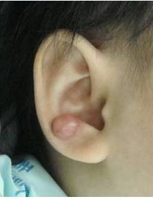
A six-month-male infant presented to a local dermatology clinic with a few weeks history of an erythematous lesion in the right ear. It was treated initially with antibiotics, but it proved to be ineffective. He was referred to dermatologists of another general hospital. Ultrasonography and fine needle aspiration biopsy (FNAB) were performed at the hospital, which were inconclusive. He was, therefore, referred to our division at 7 months of age. The mass grew rapidly in a month. Physical examination of the right antihelix demonstrated a 1.2 x 1.0 cm nodular, firm mass (Figure 1).
Figure 1: A 1.2 X 1.0 Cm Firm Mass of the Right Ear Antihelix
The overlying skin showed red discoloration. The mass was adherent to the skin and underlying tissues. Systemic and laboratory examinations were normal. There was no history of trauma. Family history was unremarkable. Benign or malignant soft tissue tumors were suspected for the tumor. Then, excisional biopsy was performed under general anesthesia. The nodule was resected together with cartilage because it was fixed to the underlying tissue. The surgical specimen revealed a subcutaneous mass measuring 0.8 x 0.8 cm. Microscopic examination demonstrated a tumor consisting of basophilic cells and eosinophilic shadow cells. Foreign body reaction was seen as well. In addition, the transition from basophilic cells to shadow cells was found (Figure 2).
Figure 2: Histopathologic Examination of the Lesion Revealed Pilomatricoma
The tumor was diagnosed as pilomatricoma. The postoperative course was uneventful (Figure 3).
Figure 3: Postoperative Appearance after One Month
Discussion
Pilomatricoma, or calcifying epithelioma of Malherbe, is a benign skin tumor that believed to differentiate towards the hair matrix and hair cortex. Kurosawa et al. studied the expression of epithelial keratin and filaggrin in pilomatricoma and suggested that pilomatricoma can differentiate towards not only hair matrix and hair cortex, but also follicular infundibulum, outer root sheath, and hair bulge (Kurosawa et al., 2009). Calcification and ossification occur in pilomatricoma. It was reported that shadow cells positive for bone morphogenetic protein (BMP)-2 may play an important part in generating bone formation in pilomatricoma (Kurosawa et al., 2000).
Though this neoplasm can present at any age, it is common in children, and more than 60% of the cases occur in the first two decades of life (Agarwal et al., 2001). The most common anatomical location for the tumor is the head and neck region, followed by the upper extremities (Lan et al., 2003). In the head and neck region, the most frequent locations are the neck, cheek, and preauricular and orbital areas. However, pilomatricomas of the ear, especially the antihelix, were rare. Pilomatricoma occurring in the ear has been observed in 0% to 4.5% of previous studies (Agarwal et al., 2001; Al-Khateeband and Hamasha, 2007; Danielson-Cohen et al., 2001; Duflo et al., 1998; Lan et al., 2003;O’Connor et al., 2011; Pirouzmanesh et al., 2003). Pilomatricomas in external auditory meatus (Vinayak et al., 1993), helix (Hassanein et al., 2011; Nassie et al., 2007), ear lobe (Rao and Ramnarayan, 1992; Pant et al., 2010; Sevin et al., 1995), and antihelix (Li et al., 2011) have been reported by several authors as rare cases.
In general, pilomatricoma is a benign, solitary, firm, and subcutaneous mass. Growth is usually slow and occurs over a period of several months or years. The overlying skin may have a reddish or bluish discoloration. Multiple occurrences have been observed in 2% to 10% of cases. A multiple nodular pattern, overlying skin erosion, and a cystic variant have been reported as well (Vinayak et al., 1993; Li et al., 2011; Duflo et al., 1998; Lan et al., 2003; O’Connor et al., 2011;Pirouzmanesh et al., 2003).
Though the presence of nodule of this type, especially in younger patients, should raise suspicion of pilomatricoma, it makes a clinical diagnosis of pilomatricoma difficult. Literature survey shows that the accuracy rate of the preoperative diagnosis of pilomatricoma ranges from 0% to 30% (Pant et al., 2010).
The differential diagnosis of pilomatricoma of head and neck should include sebaceous cyst, ossifying hematoma, brachial remnants, preauricular sinuses, adenopathy, giant cell tumor, chondroma, epidermal cyst, dermoid cyst, degenerating fibroxanthoma, foreign body reaction, osteoma cutis infantile hemangioma, parotid tumors, and malignant tumors (Lan et al., 2003; Hassanein et al., 2011; Agarwal et al., 2001; Pirouzmanesh et al., 2003; Danielson-Cohen et al., 2001; Duflo et al., 1998).
“Tent” sign and “teeter-totter” sign are the most helpful clinical findings for the diagnosis of pilomatricoma (Pant et al., 2010). “Tent” sign is produced by stretching the overlying skin to show the multifaceted components of calcification. “Teeter-totter” sign means that pressing on one edge of the lesion causes the opposite edge to protrude from the skin.
In the present case, the lesion showed atypical features clinically and it was quite difficult to make preoperative diagnosis. The tumor was located on the ear antihelix. “Tent” sign and “teeter-totter” sign were not found. The tumor has enlarged rapidly. The subcutaneous mass was adherent to the overlying skin and underlying tissues. The differential diagnoses we made have included keloid, dermatofibroma, dermatofibrosarcoma protuberans, or rhabdomyosarcoma.
When the presentation is classic, diagnosis may be made by clinical findings (Duflo et al., 1998; Pirouzmanesh et al., 2003). Radiologic imaging is of little diagnostic value for pilomatricoma (Agarwal et al., 2001; Duflo et al., 1998). Fine needle aspiration biopsy (FNAB) may be helpful. However, the results of FNAB can be misleading if there are no ghost cells present in the aspirate attributing to the misdiagnosis of many cases (Agarwal et al., 2001; Duflo et al., 1998). Excisional biopsy should be made in an atypical case as with the present case to obtain a definitive diagnosis or to alleviate symptoms ( Birman et al., 2009; Vinayak et al., 1993).
Standard treatment of pilomatricoma is surgical excision since spontaneous regression is never been observed (Agarwalet al., 2001; Duflo et al., 1998; O’Connor et al., 2011; Pirouzmanesh et al., 2003). Malignant transformation to a pilomatrix carcinoma should be suspected in cases with repeated local recurrences (van der Walt and Rohlova, 1984).
Conclusion
In conclusion, we present an infant with pilomatricoma of the ear antihelix. The pilomatricoma of the head and neck region is not uncommon, but it is rare to occur in the ear, especially the antihelix. Diagnosis of pilomatricoma may be made clinically when the presentation is classic. However, it is occasionally difficult to make diagnosis as in the present case. Excisional biopsy should be made in an atypical case to obtain a definitive diagnosis or to alleviate symptoms.
Figures and Tables
Figure 1: A 1.2 x 1.0 cm firm mass of the right ear antihelix.
Figure 2: Histopathologic examination of the lesion revealed pilomatricoma.
Figure 3: Postoperative appearance after one month.