Introduction
Cutaneous manifestations of breast carcinoma is uncommon, however, recognition of such lesions is important in establishing early diagnosis and treatment. This may be particularly difficult to identify in cases where surgical treatment and radiotherapy have been previously employed. Histopathologically, there are 8 types of cutaneous metastasis related to breast carcinoma. Here, we report a case of carcinoma en curaisse in a patient with metastatic breast carcinoma.
Case Report
A 56 year old Chinese female presented with a 7 month history of painful, discharging nodules on her chest wall. She had been diagnosed with carcinoma of the right breast (T2N2M0) in January 2011 and underwent a mastectomy with axillary lymph node clearance. Following adjuvant chemotherapy with doxorubicin, cyclophosphamide and paclitaxel, a course of 25 sessions of radiotherapy then ensued; this was completed in July 2011.Follow up imaging in December 2011 showed suspicious lesions in the lymph node and right chest wall.. In the months preceeding this consult, she had been clinically diagnosed with post radiation dermatitis and infected scars post surgery. Poor response to treatments with topical steroids and oral antibiotics prompted her request to seek a dermatological opinion.
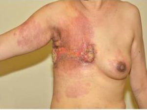
On examination, erythematous ulcerated plaques involving her right lateral and posterior chest wall was seen with tender fungating keloidal nodules. Axillary and cervical lymph nodes were palpable (fig 1).

A punch biopsy showed neoplastic cells arranged in linear cords, dissecting the collagen throughout the dermis. Immunophenotyping profile was performed and tumour cells were strongly positive for CK7 and CK8 and negative for CK20, HMB45, Melan-A and LCA (fig 2a-c).

This patient was diagnosed with recurrence of breast carcinoma, carcinoma en curaisse type and referred back to the oncologist. Imaging of the chest, abdomen and pelvis confirmed indeterminate involvement of the left axillary lymph nodes, chest wall and spleen. She has been started on Eribulin for palliative chemotherapy.
Discussion
Cutaneous metastases from solid primary tumours are uncommon and skin manifestations usually presents a few months or years following the primary diagnosis. Carcinoma of the breast is however the commonest malignancy to metastasize to skin and is usually associated with a local recurrence post therapeutic masectomy as mentioned in a papers by Mahore (2010) and Lakshmi (2010).
There are eight clinico-histopathological types of cutaneous metastasis related to breast cancer. They are carcinoma erysipelatoids, telangiectactic metastatic carcinoma, nodular metastatic carcinoma, Paget’s disease, alopecia neoplastica, carcinoma of the inframammary crease, metastatic mammary carcinoma of the eyelid with histiocytoid histology and carcinoma en cuirasse as reported in a papers by Mahore (2010), Lahore (2010), Ho (2008) and Lee (2004). A previous study by Mordenti (2000), showed the prevalence of each type of skin metastasis with nodular types being commonest (80%). Carcinoma en cuirasse was only seen in 3% of the 164 individuals in this study.
Carcinoma en cuirasse, is often likened to an encasement in armour whereby the patient presents with a diffuse indurated plaque they may be preceded by scattered, firm, papulonodules that overlie and coalescing non-inflammed erythematous plaque.
Histologically, as seen in similar cases reported by Ho (2008) and Lee (2004), it is characterized by a dense stromal fibrosis and reduced vascularity. An interstitial infiltrate of tumour cells in single rows is often seen also known as “Indian filing”. The tumour cells usually disseminate along the tissue spaces but infiltration of the lymphatic vessels may give rise to the indurated appearance. Patients with carcinoma en cuirasse do complain of burning pain as in our patient. This may be a result of perineural invasion as proposed by Lahore (2010).
As the clinical presentation is often associated with a history of previous radiotherapy, a differential of radiation therapy induced morphoea or chronic dermatitis should be considered. Other conditions which may produce similar sclerodermaotoid skin changes such as lichen sclerosus et atrophicus, graft-vs-host disease and bleomycin-induced sclerosis should also be considered. If seen earlier, this patient may have presented with nodules and hypertrophic scars more in keeping with keloid formation post surgery as described in a paper by Mullinax (2004). The erythematous plaque may also point towards an infective cause such as erysipelas.
As in the Mordenti (2004) paper, skin metastases are usually associated with advanced breast carcinoma and prognosis is poor. The dense fibrosis and decreased vascularity is thought to play a role in it chemoresistance. Therapeutic modalities that have been tried include chemotherapy, radiotherapy, hormonal antagonists and experimental combinations snake venoms as mentioned in Ho (2008) and Mullinax (2004) . In general, the prognosis is dependent on the biological behaviour and type of primary tumour. Breast carcinoma with skin metastases is usually associated with advanced cancer.
In summary, carcinoma en cuirasse is a rare form of cutaneous breast metastasis most commonly associated with local recurrence following therapeutic mastectomy. Clinicians should be aware of the various presentations of metastatic breast cancers.
Reference
Ho, M. S. L. & Tang, M. B. Y. (2008). “An Indurated Plaque on the Chest and Breasts,” Clinexpdermatol. Aug; 33 (5):665-6.
Publisher – Google Scholar
Lakshmi, C., Pillai, S. B., Sharma, C., et al. (2010). “Carcinoma En Cuirasse of the Breast with Zosteriform Metastasis,”Indian J Dermatolvenereolleprol. Mar-Apr; 76 (2):215.
Publisher – Google Scholar
Lin, J. H., Lee, J. Y., Chao, S. C., et al. (2004). “Telangiectatic Metastatic Breast Carcinoma Preceded by En Cuirasse Metastatic Breast Carcinoma,” Br J Dermatol. Aug; 151 (2):523-4.
Publisher – Google Scholar
Mahore, S. D., Bothale, K. A., Patrikar, A. D. & Joshi, A. M. (2010). “Carcinoma En Cuirasse: A Rare Presentation of Breast Cancer,” Indian J Patholmicrobiol. Apr-Jun; 53 (2):351-8.
Publisher – Google Scholar
Mordenti, C., Peris, K. M., Concetta, F., et al. (2000). ‘Cutaneous Metastatic Breast Carcinoma: A Study of 164 Patients,’ Actadermatovenerologica 2000: 9: (E-Edition).
Google Scholar
Mullinax, K. & Cohen, J. B. (2004). “Carcinoma En Cuirasse Presenting as Keloids of the Chest,” Dermatolsurg 30:226-228.
Publisher – Google Scholar


