Metastatic Small Bowel Tumours Causing Intestinal Obstruction: A Report of Two Cases

Yang-Yang Lee1, Ker-Kan Tan2 and Koh Chi-Siong Dean2

1Department of General Surgery, Singapore General Hospital, Singapore

2Division of Colorectal Surgery, National University Health System, Singapore

Academic Editor: Antonis A. Michalopoulos

Cite this Article as:

Yang-Yang Lee, Ker-Kan Tan and Koh Chi-Siong Dean (2013), "Metastatic Small Bowel Tumours Causing Intestinal Obstruction: A Report of Two Cases," International Journal of Case Reports in Medicine,
Vol. 2013 (2013), Article ID 234667, DOI: 10.5171/2013.234667

Copyright © 2013 Yang-Yang Lee, Ker-Kan Tan and Koh Chi-Siong Dean. Distributed under Creative Commons CC-BY 3.0

Abstract

Small bowel metastases arising from hematogenous seeding of primary tumours are rare. We report on two cases of subacute intestinal obstruction arising from lung and skin adnexal metastases, and review the literature on diagnostic and treatment modalities for these lesions.

Keywords: Metastasis, small bowel, subacute intestinal obstruction, skin adnexal tumour.
Introduction

Although small bowel metastases arising from primary lung cancer occur in over 10% of patients who underwent autopsy, Butler et al (1991) reports that less than 0.5% of them actually develop acute complications, such as obstruction, bleeding and perforation. Metastasis from adenocarcinoma of the skin causing small bowel complications is even rarer. In this paper, we describe two such cases and review the existing literature.

First Case Report

A 76-year-old gentleman with a chronic history of smoking, presented to our unit with complaints of colicky abdominal pain associated with bilious vomiting for five days. There was no evidence of cachexia or pallor and examination of the abdomen revealed a distended but non-tender abdomen with no evidence of peritonitis. Digital rectal examination was otherwise unremarkable.  Abdominal radiographs that were performed demonstrated dilated small bowel loops and supported the provisional diagnosis of an intestinal obstruction. A computer tomographic (CT) scan of the abdomen performed on the third day of admission demonstrated distended and oedematous small bowel loops with no obvious transition point. The patient’s symptoms resolved, and he managed to move his bowels after a further two days of nasogastric decompression and intravenous fluid hydration.

An incidental right lung nodule was noted on the routine chest radiograph. This was subsequently evaluated with a CT-guided needle biopsy which confirmed the lesion to be a poorly differentiated primary lung adenocarcinoma. Pulmonary function tests demonstrated poor pulmonary reserve and the patient was deemed a poor candidate for lung resection by the thoracic surgical team. With the apparent resolution of his obstruction, he was discharged into palliative care.

He was readmitted one day later with the similar complaints of abdominal pain with bilious vomiting. Abdominal radiographs were repeated and again highlighted dilated small bowel loops. This time, a gastrografin study was performed to determine the site of potential mechanical obstruction that might have been missed on the prior CT scan. Interestingly, the gastrografin study demonstrated good flow of contrast into the descending colon within normal transit time, with no hold up of contrast. In view of all the seemingly normal results, the patient was again managed conservatively for 9 days, before the decision was made to proceed with an exploratory laparotomy. Intra-operatively, a stenosing circumferential serosal lesion in the jejunum measuring 5.0 x 2.0 x 1.2 cm was found, with proximal bowel dilatation [Figs. 1 and 2]. A segmental small bowel resection with functional end-to-end stapled anastomosis was performed. Histological evaluation confirmed the presence of a poorly differentiated non-small cell type carcinoma extending transmurally into the serosa. Immunohistochemical staining showed immunoreactivity with CK7, P63, 35BH11, with negative staining for CK20, TTF-1, CDX-2 and D-PAS. This was consistent with a metastatic lesion from his lung primary. The patient recovered uneventfully and was discharged well on the 12th post-operative day.

234667-fig-1

Figure 1. Serosal Deposits on Small Bowel

234667-fig-2

Figure 2. Cut Specimen of the Bowel Showing Extrinsic Invasion into the Bowel Wall

 
Second Case Report

A 49-year-old lady presented with complaints of constipation and bilious vomiting for 4 days. Although she was still able to pass flatus, she was unable to even tolerate oral liquids. The patient was slightly dehydrated and her abdomen was mildly distended but not tender on palpation. She had a previous history of a periumbilical skin adnexal adenocarcinoma with resection performed. There was a periumbilical scar which was a result of surgical procedures three years ago. She had presented then with an umbilical abscess that was drained surgically. After numerous subsequent drainage procedures for a persistently recurring abscess, a CT scan was performed to further evaluate the extent of the lesion. This revealed an inflammatory mass measuring 3.1 x 4.2 cm. There was involvement of the rectus muscles and lineaalba.  She underwent a repeat debridement of the suspected abscess, and histological evaluation of the cavity wall revealed a moderately differentiated adenocarcinoma with squamous metaplasia, likely arising from the skin adnexa. The medical oncologist treated her with a course of adjuvant carboplatin and paclitaxel for ten months, followed by eight cycles of gemcitabine. Despite this, her disease continued to progress and a positron-emission tomography (PET) CT scan performed three months prior to this admission revealed a hypermetabolic mesenteric mass in the terminal ileum. She declined further chemotherapy and defaulted subsequent follow-up appointments.

An abdominal radiograph performed this time showed prominent small bowel loops with a paucity of large bowel gas shadows. The CT scan of the abdomen and pelvis demonstrated the right mesenteric abdominal mass measuring 4.4 x 3.3cm, similar to the one seen in the earlier PET-CT. However, there was now associated small bowel obstruction [Fig. 3]. She now agreed to an exploratory laparotomy which revealed two intra-abdominal lesions: a necrotic serosal mass measuring 3 cm in the proximal to mid ileum that was responsible for the intestinal obstruction; a second lesion measuring 1 cm in diameter located in the pericaecal appendices epiploicae. The loop of ileum containing the first lesion was resected, together with a limited right hemi-colectomy for the second lesion due to its proximity to the ileocaecal valve. Both functional end-to-end anastomoses were fashioned using a standard linear stapling device. The patient had an uneventful recovery and was discharged well on the 6th post-operative day. Histological examination of both lesions demonstrated moderately differentiated adenocarcinoma showing immunore activity for CK7, CK5, pCEA and CA-125, with negative staining for CK20. This was consistent to the histological findings of her previous adnexal skin cancer. Both specimens were serosal deposits with invasion down to the submucosa and muscularispropria layers.

234667-fig-3

Figure 3. Small Bowel Obstruction from Serosal Metastases to the Small Bowel


Discussion

Metastatic deposits to the small bowel are rare. Abbas (2007) and Minardi (1998) report that the majority of these lesions arise as a result of transcoelomic seeding of an intra-abdominal primary, of which colorectal cancer accounts for almost half of them. Other cancers include those of pancreatic, gastric and ovarian origins, with rarer occurrences from the lung, kidney and breast. Small bowel metastases from lung primaries are extremely uncommon, with an estimated reported incidence of 0.2-0.5% as reported in various case series by Berger (1999), Goh (2007) and Mcneill (1987). The absence of concomitant carcinomatosisperitonei in both of our cases may suggest the possible role of haematogenous seeding of tumour emboli.

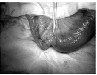
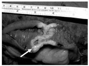
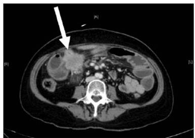
Small bowel tumours are notoriously difficult to diagnose due to their non-specific symptoms. They typically present as a surgical abdomen, with obstruction, perforation, or bleeding. These lesions usually pose significant diagnostic challenges. Facey et al (2007) reports that CT evaluation in detecting small bowel lesions has a sensitivity of 70%. It is often difficult to discern mural thickening in the presence of concomitant small bowel wall oedema, as was the case for our first patient. Some researchersrecommend capsule videoendoscopy for the assessment of the small bowel and subsequent double balloon endoscopy for biopsy of any visualized lesions. This is obviously not  possible in an acute presentation. Mosier (1992) and Facey (2007) report that PET-CT is being used more frequently in the staging of certain tumours, with the benefit of identifying early metastatic disease, as seen in the second patient.

The role of surgical intervention in the management of patients with metastases to the small bowel remains palliative in nature. While some authors such as Han (2010) and Kant (2010) advocate comfort care in patients with widespread disease, surgical intervention remains integral in patients presenting with acute surgical emergencies as seen in our patients, both of whom had a reasonable life-expectancy from localized metastatic disease. The surgical options include a palliative bypass procedure or excisional resection. Complete macroscopic clearance has been recommended in selected patients with good functional status and isolated intra-abdominal disease as this improves the quality of life and symptom-free survival. Kant (2010) reports thatlong-term survival has been reported if complete clearance of intra-abdominal disease is attained.

The overall prognosis of metastatic lung tumours to the small bowel remains abysmal, with 5-year survival rates estimated at 9-20%. In comparison, the prognosis of metastatic tumours of the skin adnexa is unknown due to its rarity, but a 5-year-survival of 42% has been reported by Blake (2010).

Conclusions

Metastases from lung and skin adnexal malignancy to small bowel causing acute complications are rare. Surgical intervention in these patients remains integral,although the long term outcome remains dismal.

References

Abbas, S. M. & Merrie, A. E. H. (2007). “Resection of Peritoneal Metastases Causing Malignant Small Bowel Obstruction,” World J SurgOnco. 5:122.
PublisherGoogle Scholar

Berger, A., Cellier, C., Daniel, C., Kron, C., Riquet, M., Barbier, J.- P., Cugnenc, P.- H. & Landi, B. (1999). “Small Bowel Metastases from Primary Carcinoma of the Lung: Clinical Findings and Outcome,” Am J Gastroenterol, 94(7) 1884-1887.
PublisherGoogle Scholar – British Library Direct

Blake, P. W., Bradford, P. T., Devesa, S. S. & Toro, J. R. (2010). “Cutaneous Appendageal Carcinoma Incidence and Survival Patterns in the United States,” Arch Dermatol. 146 (6) 625-632.
PublisherGoogle Scholar

Bulter, J. A., Cameron, B. L., Morrow, M., Kahng, K. & Tom, J. (1991). “Small Bowel Obstruction in Patients with a Prior History of Cancer,” Am J Surg, 162(6) 624-628.
PublisherGoogle Scholar

Facey, K., Bradbury, I., Laking, G. & Payne, E. (2007). “Overview of the Clinical Effectiveness of Positron Emission Tomography in Selected Cancers,” Health Technol Assess. 11 (44)  ii-iv, xi-267.
Publisher

Goh, B. K., Yeo, A. W., Koong, H. N., Ooi, L. L. & Wong, W. K. (2007). “Laparotomy for Acute Complications of Gastrointestinal Metastases from Lung Cancer: Is it a Worthwhile or Futile Effort?,” Surg Today 37 (5) 370-374.
PublisherGoogle Scholar – British Library Direct

Han, S. L., Cheng, J., Zhou, H. Z. et al. (2010). “Surgically Treated Primary Malignant Tumour of Small Bowel: A Clinical Analysis,” World J Gastroenterol. 16 (12) 1527-1532.
PublisherGoogle Scholar

Kant, K. M., Noordhoek, H. V. & Aerts, J. G. J. V. (2010). “A Patient with Four-Year Survival after Nonsmall Cell Lung Carcinoma with a Solitary Metachronous Small Bowel Metastasis,” J Oncol. 2010: 616130.
PublisherGoogle Scholar

McNeill, P. M., Wagman, L. D. & Neifeld, J. P. (1987). “Small Bowel Metastases from Primary Carcinoma of the Lung,”Cancer, 59 (8) 1486-1489.
PublisherGoogle Scholar

McNeil, P., Wagman, L. & Neifeld, J. (1987). “Small Bowel Metastases from Primary Carcinoma of the Lung,” Cancer, 59: 1486-1489.
PublisherGoogle Scholar

Minardi, A. J., Zibari, G. B., Aultman, D. F., McMillan, R. W. & McDonald, J. C. (1998). “Small Bowel Tumours,” J Am CollSurg, 186:664-668.
PublisherGoogle Scholar – British Library Direct

Mitsui, K., Tanaka, S., Yamamoto, H. et al. (2009). “Role of Double-Balloon Endoscopy in the Diagnosis of Small Bowel Tumours: The First Japanese Multicenter Study,” GastrointestEndosc. 70 (3) 498-504.
PublisherGoogle Scholar

Molina, J. R., Yang, P., Cassivi, S. D., Schild, S. E. & Adjei, A. A. (2008). “Non-Small Cell Lung Cancer: Epidemiology, Risk Factors, Treatment and Survivorship,” Mayo Clin Proc. 83 (5) 584-594.
PublisherGoogle Scholar – British Library Direct

Mosier, D. M., Bloch, R. S., Cunningham, P. L. & Dorman, S. A. (1992). “Small Bowel Metastases from Primary Lung Carcinoma: A Rarity Waiting to be Found?,” Am Surg. 58 (11)  677-682.
PublisherGoogle Scholar – British Library Direct

Trifan, A., Singeap, A. M., Cojocariu, C., Sfarti, C., Tarcoveanu, E. & Georgescu, S. (2010). “Single-Balloon Enteroscopy Following Videocapsule Endoscopy for Diagnosis of Small Bowel Tumours: Preliminary Experiences,” Chirurgia (Bucur) 105 (2) 211-217.
Publisher

 
Shares