Intraosseous Lymphoma Presenting as a Fracture of the Femoral Neck

Huan-Tee Chan1, Chi-Chou Tseng1, Po-Yih Chen1, Shih-Sung Chuang2, Wan-Lin Shen3, Chung-Da Wu4 and Wei-Ting Lin4,5

1Department of Orthopedics, Chi Mei Medical Center, Liouying, Tainan, Taiwan

2Department of pathology, Chi Mei Medical Center, Tainan, Taiwan

3Department of pathology, Chi Mei Medical Center, Liouying, Tainan, Taiwan

4Department of Orthopedics, Chi-Mei Medical Center, Tainan, Taiwan

5Department of Physical Therapy, Shu-Zen College of Medicine and Management, Taiwan

Academic Editor: Shekhar Madhukar Kumta

Cite this Article as:

Huan-Tee Chan, Chi-Chou Tseng, Po-Yih Chen, Shih-Sung Chuang, Wan-Lin Shen, Chung-Da Wu and Wei-Ting Lin (2013), "Intraosseous Lymphoma Presenting as a Fracture of the Femoral Neck," International Journal of Case Reports in Medicine, Vol. 2013 (2013), Article ID 275363, DOI: 10.5171/2013.275363

Copyright © 2013 Huan-Tee Chan, Chi-Chou Tseng, Po-Yih Chen, Shih-Sung Chuang, Wan-Lin Shen, Chung-Da Wu and Wei-Ting Lin. Distributed under Creative Commons CC-BY 3.0

Abstract

Intraosseous lymphomas are uncommon bone malignancy, and most of them present with bone pain. Herein, we describe a case of 64-year-old male who suffered from a painful swelling over his hip after a motor vehicle accident initially, and radiography showed femoral neck fracture. He received bipolar hemiarthroplasty; however, the pathological examination of surgical specimen confirmed the diagnosis of diffuse large B cell lymphoma. This demonstration should remind physicians about the possible occurrence of bone lymphoma in the clinical setting of traumatic fracture in the elderly patients.  
Keywords: Intraosseous lymphoma, femoral fracture.

Introduction

Intraosseous lymphoma can be considered as metastatic lymphoma, or as primary bone lymphoma which developed in the bone without involvement of any other site in the body. Most of the intraosseous lymphomas should be metastatic, and primary intraosseous lymphoma is a rare type of lymphoma, which accounts for only about 5% extranodal lymphoma (Pettit et al., 1990). In contrast to the classical manifestations of patients with non-bone lymphoma, symptoms, including fever, body weight losses, and night sweating, are rarely noted among patients with primary bone lymphoma. The most common presentation of the patients with intraosseous lymphoma is bone pain, and some patients have the initial presentation of a palpable mass (Qureshi et al., 2010). In addition to the nonspecific symptoms, the radiological findings of intraosseous lymphoma are quite variable and noncharacteristic. Therefore, it is not easy to make correct diagnosis initially in most clinical conditions. Herein, we described a case of unusual initial presentation of intraosseous lymphoma with traumatic femoral neck fracture.

Case Report

A 64-year-old male presented with painful swelling over right hip because of falling down from his motorcycle when riding. He denied a significant body weight loss over the preceding 6 months and any major medical diseases, such as hypertension, diabetes mellitus, and malignancy. He also did not complain of any pain or limited range of motion of right hip before the accident. His physical examination revealed ecchymosis and tenderness over right swollen hip. Pelvic X-ray showed displaced fracture at right femur (Garden stage IV) and no significant osteolytic or osteoblastic lesions (Figure 1A).

275363-fig-1

Figure 1: Radiography of Pelvis Showed Displaced Fracture at Right Femur and no Significant Osteolytic or Osteoblastic Lesions (A), and Bipolar Hemiarthroplasty was Placed and Well Fixed (B).


Bipolar hemiarthroplasty was done (Figure 1B), and there was no fragile soft tumor tissue in the bone canal.
275363-fig-1'

However, pathological examinations of right femoral head demonstrated that the bony trabeculae are intervened by fibrotic and desmoplastic marrow spaces, where sheets of necrotic atypical lymphoid cells with severe crush artifact are present (Figure 2A) and the atypical lymphoid cells are composed of medium to large cells with cleaved, hyperchromatic nuclei (Figure 2B).

275363-fig-2a

Figure 2A: The Bony Trabeculae are Intervened by Fibrotic and Desmoplastic Marrow Spaces, where Sheets of Necrotic Atypical Lymphoid Cells with Severe Crush Artifact are Present (H&E, 40X).

275363-fig-2b

Figure 2B: The Atypical Lymphoid Cells are Composed of Medium to Large Cells with Cleaved, Hyperchromatic Nuclei (H&E, 200X).


Thereafter, diffuse large B cell lymphoma with extensive necrosis was confirmed. He was discharged smoothly on postoperation day 7 and was referred to oncology outpatient department for further evaluation and management. Bone marrow aspiration was performed and revealed hypocellular marrow without abnormal lymphocytes. Computed tomography (CT) of chest and abdomen disclosed no apparent CT evidence of lymphoma lesion, but positron emission tomography (PET) of whole body displayed in favor of primary lymphoma in the right femur with other lesions probably in the spleen and interlobar lymph node (LN) of right lower lobe of lung (stage IV) (Figure 3A and 3B). So, his pathology stage was Ann Arbor stage IVA. He received chemotherapy with R-CHOP (rituximab+ cyclophosphamide + hydroxydaunorubicin + oncovin + prednisone) and had complete remission after six months. In the meanwhile, the bipolar prosthesis remained well fixed without loosening sign.

275363-fig-3
275363-fig-3'

Figure 3: Positron Emission Tomography (PET) of Whole Body Displayed in Favor of Primary Lymphoma in the Right Femur with Other Lesions Probably in the Spleen (A) and Interlobar Lymph Node (LN) of Right Lower Lobe of Lung (B) (stage IV).


Discussion

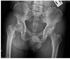
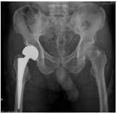
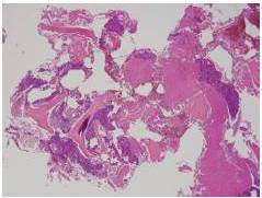
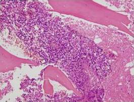
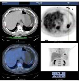
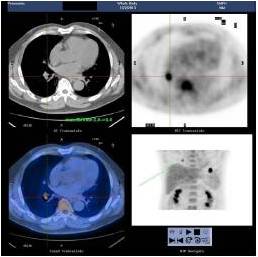
Primary lymphoma of bone is rare, and most lymphomas that involve bone are metastatic from lymph node. In this case, we demonstrated an atypical presentation of intraosseous lymphoma–traumatic femoral neck fracture in a patient. He did not present with the most common symptoms of bone lymphoma–bone pain or a mass, and his radiography of pelvis disclosed no significant bony tumor lesion, such as osteolytic, osteoblastic, and osteosclerotic signs. The final diagnosis was confirmed after pathological examinations of surgical specimens. It reminds us that the clinical manifestations of bone lymphoma are protean, even in the clinical condition of traumatic fracture.

In the recent study (Qureshi et al. 2010) that reviews a total of 60 cases of bone lymphoma during the period from 1998 to 2008, the most common site of involvement was femur (28.3%), followed by hip (16.6%) and humerus (10%). In addition, iliac bone and spine were ever reported to be involved (El-Essawy et al., 2012 and Zhu et al., 2012). This is consistent with another study by Deshmukh et al., who showed that femur (43%) was the most frequently involved site among 7 cases of bone non-Hodgkin lymphoma (Deshmukh et al., 2004). We had the similar finding that the present case had the involvement of lymphoma over his femoral neck.

Non-Hodgkin lymphoma is the most common primary bone lymphoid malignancy, and diffuse large B-cell lymphoma, such as our case, accounts for the greatest percentage of cases (Qureshi et al., 2010 and Deshmukh et al., 2004). In addition, there are several unusual cell types of bone lymphoma, such as Burkitt’s lymphoma, anaplastic large cell lymphoma, and peripheral T cell lymphoma (Qureshi et al., 2010 and Park et al., 2012). The cell type distribution may be various according to the age. In the study by Qureshi et al., the bone lymphomas of all of the 15 elderly patients with age of 60 years and above were belonged to diffuse large B cell lymphoma (Qureshi et al., 2010).

As the other type of lymphoma, the outcome of bone lymphoma was determined by the stage and cell type (Qureshi et al., 2010). In the study by Catlett et al. (2008), who investigated the survival in 30 patients with bone lymphoma, they found that the overall 5-year survival rate was 73% during a median follow-up of 49 months. Additionally, they noted a significant difference in overall survival with low and low intermediate International Prognostic Index (IPI) score versus high intermediate. In summary, patients with bone lymphoma, particularly those with low IPI scores, were shown to have good outcome and responded well to chemotherapy (Catlett et al., 2008). Overall, the treatment of these lesions may need surgery, radiotherapy, chemotherapy, or combination. In fact, lymphoma of bone usually responds well to a combination of radio- and chemotherapy regimens and can reach an overall response rate of 94% (Baar et al., 1994).

In conclusion, the clinical manifestations and radiological findings of bone lymphoma are quite variable and nonspecific. In contrast, osteoporotic (low-energy trauma) femoral neck fracture is common in the elder. Our report suggests that lymphoma can be presented as traumatic fracture due to metastatic bone lymphoma.

References

Baar, J., Burkes, R. L., Bell, R., Blackstein, M. E., Fernandes, B. & Langer, F. (1994). “Primary Non-Hodgkin’s Lymphoma of Bone. A Clinicopathologic Study,” Cancer 73(4), 1194-1199.
PublisherGoogle Scholar

Catlett, J. P., Williams, S. A., O’Connor, S. C., Krishnan, J. & Malkovska, V. (2008). “Primary Lymphoma of Bone an Institutional Experience,” Leukemia & Lymphoma 49(11), 2125-2132
PublisherGoogle Scholar

Deshmukh, C., Bakshi, A., Parikh, P., Nair, R., Pai, V., Gupta, S. et al (2004). “Primary Non Hodgkins Lymphoma of Bone a Single Institution Experience,” Medical Oncology 21(3), 263-267.
PublisherGoogle Scholar

El-Essawy, M. T., Vanhoenacker, F. M., Van, Dijck. H. & Ferrante, M. (2012). “Primary Lymphoma of Iliac Bone,” Journal Belge de Radiologie – Belgisch Tijdschrift voor Radiologi 95(6), 375.
PublisherGoogle Scholar

Park, D. A., Park, S. G. & Kim, S. W. (2012). “Solitary Lymphoblastic Lymphoma of the Thoracic Spine,” Journal of Korean Neurosurgical Society 52(6), 564-566.
PublisherGoogle Scholar
 
Pettit, C. K., Zukerberg, L. R., Gray, M. H., Ferry, J. A., Rosenberg, A. E., Harmon, D. C. et al. (1990). “Primary Lymphoma of Bone. A B Cell Neoplasm with a High Frequency of Multilobated Cells,” American Journal of Surgical Pathology 14(4), 329-334.
PublisherGoogle Scholar

Qureshi, A., Ali, A., Riaz, N. & Pervez, S. (2010). “Primary Non-Hodgkin’s Lymphoma of Bone: Experience of a Decade,”Indian Journal of Pathology Microbiology 53(2), 267-270.
PublisherGoogle Scholar

Zhu, Y. M., Ping, B. H. & Zhou, S. Y. (2012). “A Case Report of Primary Bone Lymphoma with Spinal Involvement,”Zhonghua Xue Ye Xue Za Zhi. 33(12), 1023.
PublisherGoogle Scholar

Shares