Introduction
Pseudoangiomatous stromal hyperplasia (PASH) is a benign proliferative lesion of the mammary stromal with hormonal influence.
Typical clinical presentation of PASH mimic fibroadenoma which is small unilateral painless breast lesion, discovered histologically after lumpectomy. To our knowledge, a rapidly growing bilateral huge PASH is very rare. There are only 2 cases of huge PASH that have been reported. Simple excision is usually adequate. However, in our case, diffuse and large bilateral PASH in a pregnant lady had post some difficulty in deciding the best treatment option.
Case Report
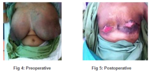
We described a case of a 40-years-old lady presented at 4 weeks of POA with bilateral breast enlargement, which was rapidly enlarged over 3 months duration. The breast enlargement had caused her severe back pain, poor appetite, and difficulty in ambulation. She has 4 children and has been taking oral contraceptive pills over the last 2 years; other than that, she has no risk factors for breast cancer. On examination, both breasts were very huge; however, there was no palpable mass. The skin appeared tense and engorged with dilated veins. No axillaries lymph nodes were palpable (Figure 4). This patient did not subject for mammogram due to very huge breasts, severe back pain, and difficulty in ambulation.
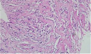
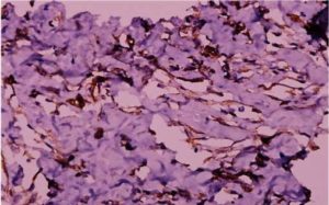
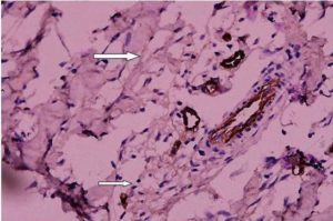
Ultrasound findings failed to locate any obvious lesion. Tru-cut biopsy was performed at the suspicious area. Histological findings revealed pseudoangiomatous stromal hyperplasia (PASH) (Figure 1). This was further supported by immunohistochemical staining that was positive for CD34 and vimentin (Figure 2) and negative for F VIII (Figure 3).

Figure 1: Pseudoangiomatous Stromal Hyperplasia Showing Abundant Stroma Containing Diffuse Slit-Like Spaces Lined by Myofibroblasts (H&E, 200 Magnification)

Fig 2: The Myofibroblasts Lining the Slit-Like Spaces are Immunoreactive for CD34 (Avidin-Biotin, 400 Magnification)

Fig 3: The Myofibroblasts are Not Immunoreactive for Factor VIII Antigen (Avidin-Biotin, 400 Magnification)
The patient was initially posted for wide local excision with hook wire localization. However, it was converted to bilateral mastectomy as the surgeon had difficulty in controlling the haemostasis. The right breast and the left breast weigh 6 and 5 kg, respectively. The HPE further confirmed the diagnosis of PASH.
Postoperatively (Figure 5), both mastectomy wounds were complicated with wound breakdown but patient’s well being successfully reestablished as she started to gain weight and to be able to ambulate as before.

Discussion
Pseudoangiomatous stromal hyperplasia (PASH) is an uncommon benign proliferative lesion of the breast, and it was first described by Vuitch et al. in 1986 (Vuitch et al., 1986; Choi et al., 2008). The clinical spectrum varies from incidental histological findings to painless unilateral breast lump. A rapidly growing bilateral huge PASH is very rare. Currently, there are only 2 cases that have been reported in the English medical literature (Ryu et al., 2010).
PASH can affect any age group especially in premenopausal women. However, male cases had also been reported recently. The exact aetiology and pathogenesis are not well understood, although PASH is thought to be associated with excessive hormonal response of mammary myofibroblast especially to the progesterone (Yo et al., 2007; Choi et al., 2008; Ryu et al.,2010; Virk and Khan, 2010).
Mammography may detect only 31% of PASH cases whereas sonographic features are nonspecific (Ryu et al., 2010).Core needle biopsy is said to have 83% sensitivity (Virk and Khan, 2010). The diagnosis is based on histological changes that characterized by complex anatomising, slit-like empty spaces in a dense fibrous stroma lined by a discontinuous layer of flat, benign spindle cells. Fine needle aspiration and cytological examination might not be helpful in diagnosing PASH (Yo et al., 2007; Choi et al., 2008; Ryu et al., 2010; Virk and Khan, 2010). Immunohistochemistry examination may be needed to support the diagnosis as the cells are positive for myofibroblastic markers including CD34, vimentin, and SMA and negative for cytokeratinin, S100, and CD31 (Virk and Khan, 2010).
PASH is not a premalignant condition and the prognosis is good. Excision of PASH with adequate margin is recommended. Recurrence had been reported up to 22% and even higher if the lesion is not completely excised (Virk and Khan, 2010). However, in our case, where patient had diffuse PASH, mastectomy had been considered as it caused significant morbidity to the patient. Although there is no established medical treatment, tamoxifen has been used in patients with small mass and breast pain (Choi et al., 2008).
Conclusion
A diagnostic delay might happen as PASH may mimic fibroadenoma clinically and its ultrasound findings were not specific. Pathologic diagnosis may be difficult unless the pathologist is aware and able to appreciate the characteristic stromal changes. Although local surgical excision is considered adequate for small localized breast mass, a generalized breast enlargement may require mastectomy in some cases.
References
Choi, Y. J., Ko, E. Y. & Kook, S. (2008). “Diagnosis of Pseudoangiomatous Stromal Hyperplasia of the Breast: Ultrasonography Findings and Different Biopsy Methods,” Yonsei Medical Journal, 49(5), 757-64.
Publisher – Google Scholar
Ryu, E. M., Whang, I. Y. & Chang, E. D. (2010). “Rapidly Growing Bilateral Pseudoangiomatous Stromal Hyperplasia of the Breast,” Korean Journal of Radiology, 11(3), 355-8.
Publisher – Google Scholar
Virk, R. K. & Khan, A. (2010). “Pseudoangiomatous Stromal Hyperplasia: An Overview,” Archives of Pathology & Laboratory Medicine, 134(7), 1070-4.
Publisher – Google Scholar
Vuitch, M. F., Rosen, P. P. & Erlandson, R. A. (1986). “Pseudoangiomatous Hyperplasia of Mammary Stroma,” Human Pathology, 17(2), 185-91.
Publisher – Google Scholar
Yoo, K.,Woo, O. H., Yong, H. S., Kim, A., Ryu, W. S., Koo, B. H. & Kang, E.- Y. (2007). “Fast Growing Pseudomatous Stromal Hyperplasia of the Breast: Report of a Case,” Surgery Today, 37, 967-970.
Publisher – Google Scholar


