Riedel’s Fibrosing Thyroiditis Associated with Elevated Serum IgG4 Levels

Amin Amraoui1, Emmanuel Rousseau2, Philippe Malvaux3 and Philippe Oriot1

1Department of Endocrinology, General Hospital of Mouscron, Mouscron, Belgium

2Department of Pathology, General Hospital of Mouscron, Mouscron, Belgium

3Department of Endocrine Surgery, Notre Dame Hospital, Tournai, Belgium

Academic Editor: Nicoletta Pedemonte

Cite this Article as:

Amin Amraoui, Emmanuel Rousseau, Philippe Malvaux and Philippe Oriot (2013), "Riedel’s Fibrosing Thyroiditis Associated with Elevated Serum IgG4 Levels," International Journal of Case Reports in Medicine, Vol. 2013 (2013), Article ID 501877, DOI: 10.5171/2013.501877

Copyright © 2013 Amin Amraoui, Emmanuel Rousseau, Philippe Malvaux and Philippe Oriot. Distributed under Creative Commons CC-BY 3.0

Abstract

Immunoglobulin G4-related sclerosing disease (IgG4-SD), also referred to as IgG4-related disease, is a newly described disease characterized by an infiltration of IgG4+ plasma cells in the early stages of the affection of an organ. Well documented in the pancreas and then detected in other organs, only few cases of thyroid affection have been reported. In addition, no case of IgG4-SD of the thyroid associated with high serum IgG4 levels has ever been described. We report here the first case.  
Keywords: Riedel’s thyroiditis; Fibrosis; IgG4-related disease.

Introduction

Fibrosis in the thyroid gland may be due to lymphoma, anaplastic carcinoma, sarcoma, multifocal sclerosing thyroiditis with lymphocyte infiltration, the sclerosing form of de Quervain’s thyroiditis, or Riedel’s disease. It has been recently shown that it could also be due to a disease associated with immunoglobulins G of the G4 subclass (IgG4), also referred to as IgG4-related disease. All these entities are remarkably similar clinically and constitute diagnostic pitfalls (Oriot, 2012; Michels, 2008). IgG4-related sclerosing disease (IgG4-SD) is a newly described fibroinflammatory disease known since 2001. It is characterized by tumors composed of dense, organized lymphoplasmacytic infiltrates rich in IgG4+ plasma cells. These lesions can progress to cause fibrosis and may be associated with an increase in serum IgG4 levels. This disease has been reported to affect many organs, including the bile ducts, salivary glands, kidneys, and lungs, as well as the pancreas, the pericardium, the periorbital tissue, the prostate, the pituitary gland, and the thyroid (Hamano, 2001). Its prevalence is currently unknown. Clinically, IgG4-SD is highly polymorphic and shows various degrees of infiltration into the organ. Thus, cases of pancreatitis jaundice, acute renal failure, or salivary swelling have been reported. To our knowledge, although the histology of the disease in the thyroid gland has been described by some authors, no case of IgG4-SD of the thyroid associated with high serum IgG4 levels has ever been described (Dahlgren, 2010; Pusztaszeri, 2012).

Case Report

A nonsmoker, asthmatic patient experienced several episodes of pleuropericarditis in 2005 at 48 years of age. A chest scan is performed, which reveals an 11x24x12 mm nodule in the right lobe of the thyroid. The thyroid-stimulating hormone (TSH) was at 1.24 μUI/ml (ranging from 0.2 to 4), and the levels of thyroid peroxidase (TPO) and thyroglobulin (Tg) antibodies were normal. A technetium-99m scintigraphy of the thyroid revealed an isofixating uptake by the nodule. Therefore, levothyroxine treatment was started in order to slow the progression of the disease. In late 2007, a total thyroidectomy was proposed, in view of the rapid growth of the nodule and its infiltrative nature on ultrasound images. During surgery, the thyroid was found to be infiltrated by whitish, sclerotic lesions, which impeded the complete removal of the gland. The histopathology revealed dense collagen bundles separating the thyroid into small lobules and the absence of granuloma. The fibrotic process extended beyond the thyroid capsule and adhered to the muscles. The cytology revealed an invasion of the thyroid by fibroinflammatory tissue composed of a polymorphic cell population of B cells, T cells, macrophages, plasma cells, neutrophils, and eosinophils (Figure 1). The diagnosis of Riedel’s thyroiditis (RT) was then established.

501877-fig-1
Figure 1: Biopsy of Thyroidectomy from 2007 Stained with Hematoxylin Erythrosin Saffron (HES) Showing Significant Fibrosis of the Thyroid Associated with a Plasma Cell Dominated Inflammatory Infiltrate. Some Lymphohistiocytic and Eosinophilic Elements Can also be Observed.
 
Several weeks later, the patient was hospitalized for severe abdominal pain in the right flank. Renal colic was then suspected. A computed tomography (CT) was performed, which revealed a flow of fibrotic tissue into the right cervical and paramediastinal area extending up to the retroperitoneal space and sheathing the thoracoabdominal aorta and right renal vessels. Corticosteroid treatment with methylprednisolone (0.5 mg/kg/day) was started and continued for nine months and then decreased to 10 mg/day (Vaidya, 1997), resulting in a significant regression of the fibrosis (Figure 2).

501877-fig-2
Figure 2: Regression of Retroperitoneal Fibrosis after 9 Months of Corticosteroid Therapy
 
In order to avoid the deleterious effects of corticosteroid treatment, tamoxifen (20 mg twice daily) was also prescribed for its antifibrotic properties (Perimenis, 2008). Thyroid laboratory results, as well as phosphate and calcium levels, remained normal without substitution. In 2009, the metabolic activity of the fibroinflammatory tissue had disappeared, as assessed by (18)FDG-PET/CT, and the bitherapy could then be stopped after 12 months of treatment.

A year later, in August 2010, a new (18)FDG-PET/CT was performed, which showed the appearance of hypermetabolic tissue at the apex of the left lung and at a the level of a mediastinal flow located around the descending thoracic aorta, with the latter showing a significant hypermetabolism (SUVmax = 10). Treatment with tamoxifen was resumed as monotherapy until late 2011; however, RT progressed into a neck mass of 4 cm in diameter located at the left lower carotid-jugular region (Figure 3).

501877-fig-3
Figure 3: Left Neck Mass of 4 cm in Diameter Revealed by the (18) FDGPET/CT and Located at the Height of the Inferior Carotid-Jugular Region.
 
This mass then caused progressive dyspnea and stridor associated with laryngeal nerve palsy causing paresis of the left vocal cord in adduction. A laser posterior cordectomy was then performed to free up space for ventilation, resulting in a rapid improvement in the respiratory condition of the patient. Corticosteroid treatment (1 mg/kg/day) was started again and a biopsy of the mass was performed three weeks after treatment onset. The histopathology revealed a dissection of adipose and muscle tissues by dense, pauci-inflammatory fibrosis. Total serum IgG levels were 1,952 mg/dL (ranging from 800 to 1,700), and serum IgG4 levels were particularly high at 333 mg/dL (ranging from 1 to 104), as determined by immunonephelometry.

The diagnosis of IgG4-related disease of the thyroid was then suspected.

Discussion

RT is very rare. At Mayo Clinic, 21 cases have been identified between 1976 and 2008 (Fatourechi, 2011; Hennessey, 2011). Histologically, RT is characterized by lymphoplasmacytic infiltrations associated with local and systemic fibrosis (Papi, 2004). It can be suspected in patients with a rapidly progressive, indurated goiter. The evolution of the disease can be marked by the onset of symptoms of tracheal compression with stridor, dysphonia due to laryngeal nerve damage, or superior vena cava syndrome. In time, retroperitoneal and/or mediastinal fibrosis, orbital pseudotumors, sclerosing cholangitis, and polyserositis can also develop in patients with RT (Perimenis, 2008; Fatourechi, 2011; Hennessey, 2011; Papi, 2004; Erdogan, 2009; Yasmeen, 2002).

The IgG4-related syndrome was first described in Japan, in the form of the so-called autoimmune pancreatitis (AIP), the main histological and serum marker of which is IgG4. IgG4 is the smallest of the four subclasses of IgGs (IgG1, IgG2, IgG3, and IgG4). The IgG1 subclass constitutes nearly 70% of all IgGs, while the IgG4 subclass constitutes less than 5% of the total pool of IgGs. All subclasses play a major role in defense against infection, by taking part in the process of opsonization and activation of the complement. However, IgG4s do not have any activity on the complement and play a limited role in autoimmunity, or their role has not yet been identified (Kakudo, 2012).

The link between IgG4-related disease and RT has already been proposed in 2010 by Dahlgren et al., who reported three cases of RT and noted common features between this disease and IgG4-SD (Dahlgren, 2010). More recently, in 2012, Pusztaszeri et al. reported a case of IgG4-SD of the thyroid, identified at first as RT (Pusztaszeri,2012). However, no case of IgG4-SD of the thyroid associated with high serum IgG4 levels has ever been described. Recently, some authors also suggested a connection between IgG4-SD and Hashimoto’s thyroiditis, which is characterized by a lymphoplasmacytic infiltration, fibrosis, and an increase in the number of IgG4+ plasma cells in the thyroid (John, 2012).

Histologically, IgG4-SD is also characterized by the presence of dense, lymphoplasmacytic infiltrates (mostly T cells), vessel occlusions by microthrombi, moderate infiltrations of eosinophils, and the formation of a dense, fibrous mass dissecting the organ. In terms of immunity, it seems that the disease is mainly mediated by Th2 lymphocytes, which are the predominant elements in the affected sites. An increase in the levels of interleukins 4, 5, 10, and 13 can also be noted in affected tissues. These cytokines induce hypereosinophilia and an increase in IgE levels in approximately 40% of patients. They also induce a proliferation of B cells, which mature into IgG4-secreting plasma cells. In addition, an activation of Treg cells can be observed, which induces the hypersecretion of TGF-β at the origin of the phenomenon of fibrosis observed in this syndrome. However, the mechanism responsible for the specific overexpression of IgG4s has not yet been elucidated (Kakudo, 2012).

The main problem is the lack of validated criteria for the diagnosis of the disease. Most criteria proposed in the literature concern AIP. Here, we used the following criteria: (i) diffuse hypertrophy of an organ; (ii) serum IgG4 levels higher than 135 mg/dL; and (iii) histopathological findings including a lymphoplasmacytic infiltration and fibrosis rich in IgG4+ plasma cells, with a plasma IgG4s/IgGs ratio greater than 40%—50%, or an infiltration of IgG4+ plasma cells >10 by large field. The diagnosis of IgG4-related disease is established in patients with two criteria or the sole presence of the third criterion. Moreover, other conditions, such as neoplasia, lymphoma, and diseases related to IgG4-SD (Sjögren’s syndrome, sclerosing cholangitis, etc.), should be excluded (Kakudo, 2012; Ebbo, 2012).

IgG4 levels can also be normal (30% of AIP) and the tissue density in IgG4+ plasma cells can be low, if prednisone is administrated before the analyses are performed. IgG4-SD is known to be sensitive to cortisone, as perfectly shown in the present case. Thus, the patient had IgG4 levels of 333 mg/dL at the time of the recurrence, but three weeks after the start of corticosteroid therapy and after the biopsy of the jugular mass, the levels of IgG4s in serum dropped to 86 mg/dL. The histology revealed mostly dense, fibrous, and pauci-inflammatory tissue, and it was impossible to quantify the number of IgG4+ plasma cells. To confirm the hypothesis of IgG4-SD in this patient, additional histological analyses were performed on the biopsies of the thyroid, prior to treatment with corticosteroids in 2007.

Specific immunostainings revealed an increase in IgG+ plasma cells, especially IgG4+ plasma cells, with >10 IgG4+ plasma cells by large field of 0.6 mm in diameter (Illustration 1).

Illustration 1: Cytology and IgG- and IgG4-specific immunostainings of thyroid biopsies from 2007 (photographs 1 and 2) and of the left paravertebral mass from 2012 (photograph 3).

501877-photo-1
Photograph 1: Immunoperoxidase Staining of IgGs Resulting in a Brown Staining of Numerous Plasma Cells.

501877-photo-2
Photograph 2: Immunoperoxidase Staining of IgG4s: 12-14 IgG4+ Plasma Cells per Large Field of 0.6 mm of Diameter.

501877-photo-3

Photograph 3: Staining with Hematoxylin Erythrosin Saffron (HES) Showing a Very Weak Inflammation of the Fibrous Tissue.

 
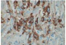
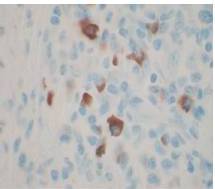
Therefore, the high levels of IgG4+ plasma cells at the time of the recurrence, the number of plasma cells expressing IgG4 > 10, the absence of neoplastic processes, such as carcinoma, lymphoma, or sarcoma, the absence of TPO and Tg antibodies, the breaking of the thyroid capsule, the regression of the tumor, and the decrease in the levels of circulating IgG4s under corticosteroids enabled us to establish the diagnosis of IgG4-related syndrome of the thyroid. Moreover, the (18)FDG-PET/CT did not reveal any other organ affection due to the disease. After six months of treatment with methylprednisolone, with a dose reduction over time, the disease did not progress anymore and IgG4 levels remained within the normal range. At six months, the ultrasound showed a significant decrease of the carotid-jugular mass.

The contribution of IgG4 levels in the diagnosis of IgG4-SD, which shows a sensitivity of 71% to 93% for AIP, is highly debated in the literature. A recent study showed that a threshold of IgG4 levels at 140 mg/dL had a sensitivity of 76%, a specificity of 93%, and positive predictive value of 36%. These rates were 53%, 99%, and 75% with a double threshold of 280 mg/dL for AIP (Lamia, 2011). To date, no data is available for the thyroid, but IgG4 levels of 333 mg/dL in our patient enabled us to confirm the diagnosis. However, IgG4 levels should be cautiously interpreted, depending on the clinical, histopathological, and immunohistochemical context and current or previous treatments administered. Carbonic anhydrase (CA) and lactoferrin antibodies can be found in most patients with AIP (16), antinuclear antibodies (ANAs) can be found in 43% to 75% of cases, and rheumatoid factor (RF) can be found in 13% to 30% of cases (Lamia, 2011). Here, antibodies to CA, RF, and ANA could not be found.

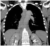
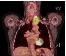
In the present study, we also confirm that (18)FDG-PET/CT provides valuable data for the monitoring of IgG4-SD. It constitutes a test of choice to assess the extent and metabolism of IgG4-SD (Nguyen, 2011). In addition, although we used tamoxifen for its antifibrotic effects, monotherapy with tamoxifen for more than 12 months did not allow for the remission of IgG4-SD, unlike the case in the study by Chacko et al. (Chacko, 2009).

In conclusion, significant similarities exist between RT and IgG4-SD, such as the presence of invasive fibrosis, lymphoplasmacytic infiltration, and good response to steroid therapy, which have been often reported and are well illustrated in the present case. RT associated with a high number of IgG4+ plasma cells, with or without elevated serum IgG4 levels, may constitute the first symptoms of a case of IgG4-SD. The assessment of a patient with thyroid fibrosis should ideally include an immunohistochemical evaluation of plasma cells (IgG+ and IgG4+ cells) and of circulating levels of IgG4s. Treatment with corticosteroids seems to be the cornerstone. It favorably influences the course of the disease. The monitoring of circulating IgG4s in the patient under treatment with corticosteroids correlated with a regression of the tumor in the present case. In addition, the recurrence of the disease in the patient under tamoxifen monotherapy is challenging. Therefore, more data is needed to clarify the link or the difference between these two entities.

References

Chacko, S., et al. (2009). “Treatment of Hyper-IgG4 Disease with Sequential Corticosteroids and Tamoxifen – Case Report and Review of the Literature,” Clinical Nephrology, 72:414-7.
PublisherGoogle Scholar

Dahlgren, M., Khosroshahi, A., Nielsen, G. P., Deshpande, V. & Stone, J. H. (2010). “Riedel’s Thyroiditis and Multifocal Fibrosclerosis are Part of the IgG4-Related Systemic Disease Spectrum,” Arthritis Care & Research (Hoboken), 62:1312-8.
PublisherGoogle Scholar

Ebbo, M., Grados, A., Daniel, L., Vély, F., Harlé, J. R., Pavic, M. & Schleinitz, N. (2012). “IgG4-Related Systemic Disease: Emergence of a New Systemic Disease? Literature Review,” La Revue de Medecine Interne, 33:23-34.
PublisherGoogle Scholar

Erdoğan, M. F., Anil, C., Türkçapar, N., Ozkaramanli, D., Sak, S. D. & Erdoğan, G. ( 2009). “A Case of Riedel’s Thyroiditis with Pleural and Pericardial Effusions,” Endocrine, 35:297-301.
PublisherGoogle Scholar

Fatourechi, M. M., Hay, I. D., McIver, B., Sebo, T. J. & Fatourechi, V. (2011). “Invasive Fibrous Thyroiditis (Riedel Thyroiditis): The Mayo Clinic Experience, 1976-2008,” Thyroid, 21:765-72.
PublisherGoogle Scholar

Hamano, H., Kawa, S., Horiuchi, A., Unno, H., Furuya, N., et al. (2001). “High Serum IgG4 Concentrations in Patients with Sclerosing Pancreatitis,” The New England Journal of Medicine, 344:732-8.
PublisherGoogle Scholar

Hennessey, J. V. (2011). “Clinical Review: Riedel’s Thyroiditis: A Clinical Review,” The Journal of Clinical Endocrinology & Metabolism, 96:3031-41.
PublisherGoogle Scholar

Kakudo, K., Li, Y., Taniguchi, E., Mori, I., Ozaki, T., Nishihara, E., Matsuzuka, F. & Miyauchi, A. (2012). “IgG4-Related Disease of the Thyroid Glands,” Endocrine Journal, 59:273-81.
PublisherGoogle Scholar

Kallel, L., Naija, N., Boubaker, J. & Filali, A. (2011). “La Pancréatite Auto Immune: Revue Systématique de la littérature,”La tunisie Medicale, 89:221-30.
PublisherGoogle Scholar

Michels, J.- J. & Blanc-Fournier, C. (2008). “What Diagnosis Can be Made by the Pathologist in a Case of Sclerosing Thyroiditis?,” Annales de Pathologie, 28:263-7.
PublisherGoogle Scholar

Nguyen, V. X., De Petris, G. & Nguyen, B. D. (2011). “Usefulness of PET/CT Imaging in Systemic IgG4-Related Sclerosing Disease. A Report of Three Cases,” JOP. Journal of the Pancreas, 12:297-305.
PublisherGoogle Scholar

Oriot, P., Malvaux, P., Waignein, F., Delcourt, A., Doyen, C., Rousseau, E., Baudry, G. & Dechambre, S. (2012). “Nodular Sclerosing Hodgkin’s Disease Mimicking Riedel’s Invasive Fibrous Thyroiditis,” Annales d’Endocrinologie (Paris),73(5) 492-6.
PublisherGoogle Scholar

Papi, G. & LiVolsi, V. A. (2004). “Current Concepts on Riedel Thyroiditis,” American Journal of Clinical Pathology, 121:50-63.
PublisherGoogle Scholar

Perimenis, P., Marcelli, S., Leteurtre, E., Vantyghem, M. C. & Wémeau, J. L. (2008). “Riedel’s Thyroiditis: Current Aspects,” Presse Medicale, 37:1015-21.
PublisherGoogle Scholar

Pusztaszeri, M., et al .(2012). “Riedel’s Thyroiditis with Increased IgG4 Plasma Cells: Evidence for an Underlying IgG4 Related Sclerosing Disease?,” Thyroid, 22 (9):964-8.
PublisherGoogle Scholar

Stone, J. H., Zen, Y. & Deshpande, V. (2012). “IgG4-Related Disease,” The New England Journal of Medicine, 366:539-551.
PublisherGoogle Scholar

Vaidya, B., Harris, P. E., Barrett, P. & Kendall-Taylor, P. (1997). “Corticosteroid Therapy in Riedel’s Thyroiditis,”Postgraduate Medical Journal, 73:817-9.
PublisherGoogle Scholar

Yasmeen, T., Khan, S., Patel, S. G., Reeves, W. A., Gonsch, F. A., de Bustros, A. & Kaplan, E. L. (2002). “Clinical Case Seminar: Riedel’s Thyroiditis: Report of a Case Complicated by Spontaneous Hypoparathyroidism, Recurrent Laryngeal Nerve Injury, and Horner’s Syndrome,” The Journal of Clinical Endocrinology & Metabolism, 87:3543-7.
PublisherGoogle Scholar

Shares