Makram Koubaa, Abir Aouam, Adnene Toumi, Chawki Loussaief, Hajer Ben Brahim and Mohamed Chakroun
Depatment of Infectious Diseases, Fattouma Bourguiba University Hospital, Monastir, Tunisia
Volume 2013 (2013),
Article ID 507024,
International Journal of Case Reports in Medicine,
4 pages,
DOI: 10.5171/2013.507024
Received date: 18 March 2013; Accepted date: 10 April 2013; Published date: 22 May 2013
Academic Editor: Héla Elloumi
Cite this Article as:
Makram Koubaa, Abir Aouam, Adnene Toumi, Chawki Loussaief, Hajer Ben Brahim and Mohamed Chakroun (2013), "Psoas Abscess as the First Clinical Manifestation of Crohn’s Disease," International Journal of Case Reports in Medicine, Vol. 2013 (2013), Article ID 507024, DOI: 10.5171/2013.507024
Psoas abscess is rare as the first clinical manifestation of Crohn’s disease. We report a 28 year-old man with no previous history of gastrointestinal disorder, who presented with lower right abdominal pain and psoitis. Diagnosis of psoas abscess was confirmed by computed tomography. Extended-spectrum beta-lactamase producing Escherichia coli were isolated from abscess. Colonoscopy and histological examination of the biopsies established the diagnosis of Crohn’s disease. Following the administration of antibiotics and computed tomography-guided percutaneous drainage, a right hemicolectomy and iliectomy resection were performed. Clinician should have a high index of suspicion to make promptly the diagnosis of Crohn’s disease presented with psoas abscess as the first manifestation especially when gastrointestinal symptoms may not dominate the complaints.
Psoas abscess (PA) remains a rare condition characterized by an insidious onset. The diagnosis is frequently delayed due to its variable and non specific clinical features. Abscesses caused by hematogenous dissemination of bacteria are defined as primary abscesses (Shields et al., 2012). In secondary PA, infection is spread by direct contact to an infectious focus frequently involving kidney, spine or gut. Crohn’s disease (CD) is most commonly associated with secondary PA (Ogihara et al., 2000). We report a case of PA revealing CD.
Case Report
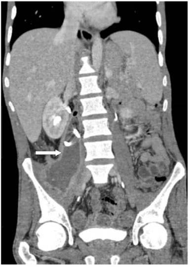
A 28-year-old man with history of appendectomy was admitted for investigation of right lower abdominal and hip pain associated with mild fever during the last two months. He was without occupation and he had no contact with other patient. There was no history of nausea, vomiting, or change in bowel habit but he had lost 7 kg over the six months before admission. He didn’t receive antibiotics during the last 6 months. The hip pain was initially intermittent, and then constant at rest, making ambulation difficult. Physical examination on admission revealed tenderness of the right lower abdomen and flexion contracture of the right hip joint. His temperature was 38.5°C. Laboratory findings were as follows: hemoglobin: 9.4 g/dL, white blood cell count: 16.500/L, erythrocyte sedimentation rate: 52 mm/h and C-reactive protein: 190 mg/L. Human immunodeï¬ciency virus serology was negative. No bacteria were isolated from the cyto-bacteriological analysis of the urine or blood culture. Computed tomography (CT) scan of the abdomen and pelvis showed a large right PA (160 x 30 mm) (Fig. 1) and severe inflammation of the terminal ileum, cecum, and ascending colon without spinal involvement. CT-guided percutaneous drainage of the abscess was performed. Aspiration revealed a purulent fluid with isolates Extended-Spectrum Beta-Lactamases (ESBL) producing Escherichia coli (resistant also to quinolones and amino-glycosides). Ertapenem was given intravenously at 1 g daily, and the patient was kept on total parenteral nutrition. Crohn’s ileocolitis was conï¬rmed by colonoscopy with biopsy. Histological examination showed no sign for tuberculosis. We suspected the presence of a fistula from the cecum to the right psoas muscle, which was confirmed by a gastrografï¬n enema. Repeat CT at two weeks showed complete disappearance of the PA. At laparotomy, the terminal ileum, right colon, and proximal third of the transverse colon were grossly diseased with a colo-colic, retrocolic and colo-ileal ï¬stulae. A right hemicolectomy and iliectomy with ileo-colic anastomosis was carried out with favorable outcome. Our patient received prophylactic anti-coagulation with low-molecular-weight heparins either pre- and postoperatively. He was discharged on oral antibiotics, steroids, and azathioprine. Four months follow-up revealed a 3-kg weight gain and resolution of symptoms on maintenance azathioprine without side effects.
Figure 1. Frontal Reconstruction of Computed Tomography Abdominal Scan Showing Right Psoas Abscess with Percutaneously Placed Drainage Catheter (Arrow)
Discussion
Intra-abdominal abscesses have been reported to occur in 6.8 to 28% of patients with CD. However, the incidence of PA is much lower (0.4—4.3%) (Jawhari et al., 1998). PA due to CD is the result of direct contact of any fissure in ulcerated intestine which penetrates backwards, on the right from the terminal ileum or cecum and on the left from jejunum or sigmoid colon. Clinical presentation is variable and diagnosis may be delayed due to the insidious onset of symptoms. The classic triad of fever, pain (in the hip, back, groin or abdomen) and limp or hip contracture is found only in 8% of cases. The diagnosis may be suspected if patient had supine position, with the knee moderately flexed and the hip mildly externally rotated. The physical examination is crucial for prompt PA diagnosis. Laboratory tests are helpful as they reveal increased white cell count, anemia, and elevated sedimentation rate. Blood cultures may be positive for a particular organism causing the abscess. Enteric bacteria (Escherichia coli, Enterobacter and Salmonella) predominate in secondary PA. CT abdominal scan with contrast is the most efficient and accurate imaging study in diagnosing a PA. It may reveal also any coexisting retro or intra-peritoneal disease as the etiology. The presence of gas bubbles or air fluid level is considered more specific in PA. Retroperitoneal magnetic resonance imaging is complex, however it may contribute to diagnose complicating small bowel CD and other sources of primary disease. Intravenous urography may be useful to diagnose possible compressions of the urinary tract. Ultrasonography lead to diagnosis in only 60% of cases and is influenced by gaseous bowel distension. Barium enema may be indicated when a complete colonoscopy cannot be performed for technical reasons or to rule out bowel origin. Management of PA complicating CD remains controversial. CT-guided percutaneous drainage and antibiotic therapy may be tried initially. Percutaneous abscess drainage in CD is now accepted as a fundamental pre-operative temporizing procedure to improve the patient’s condition before surgery (Golfieri et al., 2006). It decreases the patient’s catabolism and improves consequently nutritional status through rapid control of sepsis. The operative field can then be converted to a non infected area, thus raising the odds of safe healing after a single operation for resection of a diseased bowel with immediate anastomosis, rather than resorting to the standard two-stage procedure (Muller-Wille et al., 2011). Emergence and dissemination of ESBL producing Escherichia coli in the community has become a worldwide problem. Delayed recognition and inappropriate antibiotics of severe infections caused by ESBL producers with cephalosporin has been associated with increased mortality. Our patient didn’t receive any antibiotics six months before admission however we isolated ESBL producing Escherichia coli in the PA. Recurrent abscesses after drainage are reported in 2—8% of cases and related to the presence of fistulas (Gervais et al., 2004). Our case underwent subsequent surgery because of his fistula. The mortality from PA complicating CD has been reported to 10% (Ricci and Meyer, 1985).
Conclusion
PA may rarely reveal a CD. The diagnosis is challenging because of non-specific clinical features and gastrointestinal symptoms may not dominate the complaints. Drainage of the abscess and resection of the fistula and the affected bowel segment is the therapy of choice in these cases. Physician should be aware about the presence of ESBL in out-patient and use an appropriate antibiotics.