Discussions
Presacral Haemorrhage is bleeding resulting from injury to the presacral venous plexus during the dissection of the pelvic viscera from the sacrum. Presacral fascia is a protective layer on top interlinking thin-walled venous plexus that lines the periosteum of the sacral bone(Wydra et al., 2004). Presacral fascia is minimally vascular, hence, the haemostasis is usually easy to control; however, should there be dissection deeper and away from this fascia, the presacral venous plexus would be injured and bleeding would be torrential.
The presacral haemorrhage is rare; however, if it happens, it is lifethreatening matter that leads to shock and death. This is commonly encountered in pelvic surgery especially rectal surgery and gynaecological surgery. Stylianos et al. reported the incidence of severe presacral haemorrhage of 3.6%, and according to their literature review, there has been reported incidence of 3% to 9.4%. Unfortunately, we have no local data yet to compare(Germanos et al., 2010).In the era of laparoscopic surgery, this occurrence would become even rarer due to the magnification of the surgical field by the laparoscope whose resection is done under vision; hence, injury to the nerve and also the veins can be avoided (Arnaud et al., 2000).
Generally, when one encounters bleeding, we would put pressure compression. This rule is applicable to presacral venous haemorrhage. Conventionally, there are several ways used to achieve haemostasis such as cautery, suture ligation, hot packs, ligation of internal iliac artery, and application of absorbable gelatin sponge or microfibrillar collagen (Ayuste E, 2004 and Kumar et al., 2007).
The more widely acceptable techniques previously were pelvic packing and reexploration. There has been a report on the effectiveness of solely pelvic packing to achieve haemostasis in presacral venous haemorrhage. Wydra et al. described the success of surgical pelvic packing as a means of controllingmassive intraoperative bleeding during pelvicposterior exenteration in their case report. This technique was adopted initially from Pringle when he described liver packing for haemostasis of hepatic injury in 1908. This technique was then modified further by Halsted in 1913. In 1926, Logothetopulos first described in full extent the ways of pelvic packing for massive haemorrhage with gauge. His technique was further improvised by Parente in 1962, by replacing the gauge veil with polyethylene sheet for packing to achieve less adhesions and to facilitate the removal of the pelvic pack during reexploration (Wydra et al., 2004).
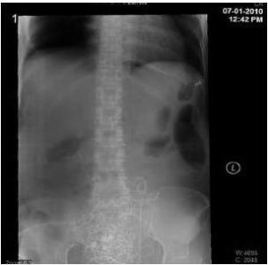
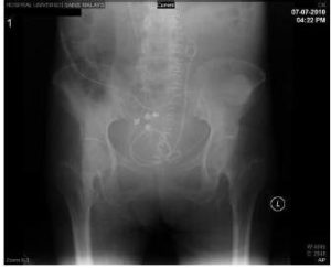
In our case, in view of the situation, we proceed with ligation of the bilateral internal iliac artery in the attempt to reduce the blood pooling at the pelvic cavity. However, both of our attempts were unsuccessful. Whilst waiting for the effect of the ligation, the surgeon in charge ordered for autoclaving of the normal thumbtacks available. Just as what we have anticipated, the bleeding does not stop despite all the described effort. The thumbtacks were picked up with a forceps, the bleeding sites at the sacrum were identified, and the thumbtacks were applied on top of the bleeders presuming that it is the foramina from which the basivertebral veins perforated through. After four thumbtacks were instilled, the ooze finally slows down.
Furthermore, we packed the pelvic cavity with six roller gauges bound together along with their radio opaque indicator and removed under local anaesthesia plus analgesic.
The application of thumbtack for haemostasis of the presacral venous haemorrhage was first described in the literature by Wang et al. in 1985 (Wang et al., 1985). They introduced the new concept to the causative injury causing the torrential bleeding at the presacral venous plexus. They noticed that there are several large foramina emerging at the lower sacral bone in the study specimens. They postulated that the basivertebral veins which emerge from these foramina would tend to bleed torrentially because of their valveless nature and also due to the retraction of the vessels into the sacral bone during resection, making cauterization or suture ligation almost impossible. Hence, they introduced the usage of thumbtacks on these bleeding sites for the control of haemostasis.
Arnaud et al. have introduced a malleable applicator designed for precise positioning of thumbtacks within the pelvis. They suggested that there should be sterile titanium thumbtacks available in all operating room where abdominoperineal resection and resection of the prolapsed rectum are performed, in anticipation of presacral haemorrhage (Arnaud et al., 2000).
Further works were done on this issue by Stolfi et al. with introduction of different pin occluder’s lengths at the sacrum in substitution of the conventional titanium thumbtacks (fixed length); they used 12 mm shaft-pins at S1 and S2 whilst 7mm shaft-pins at S3 to S5. This is an adaptation to the thumbtacks in accordance with the anatomy and the depth of the sacral bone (Stolfi et al., 1992).
We understand that thumbtacks are a foreign body; therefore, it would cause local tissue irritation leading to adhesions and it may also act as septic foci. When it is used for haemostasis, it is usually permanent and would not be removed. There has been a report on thumbtacks causing anastomosis dehiscence and septic shock, whereby removal was performed via a rigid cystoscope (Mahmalji et al., 2009).
Thumbtacks are sharp; if improperly placed, they may be dislodged and cause injury to the surrounding viscera. However, there are not many reports on them being dislodged and causing secondary injury. Suh et al. reported on a failure of the thumbtacks as the last resort to secure haemostasis in a gunshot injury to the presacral venous plexus. They postulated that these dislodged thumbtacks could have possibly caused secondary injury to the viscera and solid organs; however, they were not able to prove it. They also suggested extra precautions on the thumbtack use as it would pose a surgical hazard to both the pathologist and the surgeon, should postmortem or reexploration of the pelvic cavity be required (Suh et al., 1992)
There are other suggested ways of securing presacral haemorrhage should the thumbtacks be unavailable or should the surgeons be not comfortable in using these sharp foreign objects. The usage of local haemostatic agents such as the matrix haemostatic sealant and absorbable haemostat is also described in the literature. Matrix haemostatic sealant consists of a gelatin matrix with a granular physical structure and a thrombin component.It is applicable to hard and soft tissues as well as actively bleeding sites. The gelatin matrix will act as a coating while the mixed one along thrombin component will serve as a coagulant, hence, the action of a sealant (Germanos et al., 2010).
The absorbable haemostat is a knitted fabricprepared by oxidizing regenerated cellulose with a consistency similar to that of cotton. Theoretical proposition of haemostasis is through several mechanisms, namely, blood absorption, followed by surface interaction with proteins and platelets with activation of both the intrinsic and extrinsic pathways(Germanos et al., 2010).
Wang et al. in their 2009 case report described the successful tamponade of presacral haemorrhage with a titanium table fixation staple and a cancellous bone graft
fixed to the sacrum(Wang et al., 2009).
Muscle welding technique has been first described in the literature by Xu and Lin in 1994; in 2003, Harrisons et al. reported their experience with the technique; this is further validated by Ayuste et al. in 2004 (Harrison et al., 2003 and Ayuste E, 2004).This is performed by excising a small piece of rectus muscle, holding it to a forceps, and pressing against or close to the bleeding sites, followed by electrocautery of the forceps to char the muscle piece and weld on to the bleeding sites. This process can be repeated several times until haemostasis is secured (Ayuste E, 2004).
References
Arnaud, J.- P., Tuech, J.- J. & Pessaux, P. (2000). “Management of Presacral Venous Bleeding with the Use of Thumbtacks,” Digestive Surgery,17 651-652.Publisher – Google Scholar
Ayuste, E. & Roxas, M. F. T. (2004). “Validating the Use of Rectus Muscle Fragment Welding to Control Presacral Bleeding during Rectal Mobilization,” Asian Journal of Surgery 27 (1), 18-21.
Publisher – Google Scholar
Germanos, S., Bolanis, I., Saedon, M. & Baratsis, S. (2010). “How I Do it: Control of Presacral Venous Bleeding during Rectal Surgery,” The American Journal of Surgery 200 e33-e35.
Publisher – Google Scholar
Harrison, J. L., Hooks, V. H., Pearl, R. K., Cheape, J. D., Lawrence, M. A., Orsay, C. P. & Abcarian, H. (2003). “Muscle Fragment Welding for Control of Massive Presacral Bleeding during Rectal Mobilization: A Review of Eight Cases,”Diseases of Colon & Rectum,46 (8), 1115-1117.
Publisher – Google Scholar
Kumar, S., Malhotra, N., Chumber, S., Gupta, P., Aruna, J., Roy, K. K. & Sharma, J. B. (2007). “Case Report: Control of Presacral Venous Bleeding, Using Thumbtacks,” Archives of Gynecology and Obstetrics 276 386-386.
Publisher – Google Scholar
Mahmalji, W., Mackenzie, H., Chopada, A. & Raza, A. (2009). “Case Report: A Novel Use for the Rigid Cystoscope: The Removal of Sacral Tacks after a Coloanal Anastamosis Dehiscence,” Advances in Urology, 1-3.
Publisher – Google Scholar
Qinyao, W., Weijin, S., Youren, Z., Wenqing, Z. & Zhengrui, H. (1985). “New Concepts in Severe Presacral Hemorrhage during Proctectomy,” Archives of Surgery,120 1013-1020.
Publisher – Google Scholar
Stolfi, V. M., Milsom, J. W., Lavery, I. C., Oakley, J. R., Church, J. M. & Fazio, V. W. (1992). “Newly Designed Occluder Pin for Presacral Hemorrhage,” Diseases of the Colon & Rectum 35 (2), 166-169.
Publisher – Google Scholar
Suh, M., Shaikh, J. R., Dixon, A. M. & Smialek, J. E. (1992). “Failure of Thumbtacks Used in Control of Presacral hemorrhage,” The American Journal of Forensic Medicine and Pathology,13 (4), 324-325.
Publisher – Google Scholar
Wang, L.- T., Feng. C.- C., Wu, C.- C., Hsiao, C.- W., Weng, P.- W. & Jao, S.- W. (2009). “The Use of Table Fixation Staples to Control Massive Presacral Hemorrhage: A Successful Alternative Treatment. Report of a Case,” Diseases of the Colon & Rectum,52 (1), 159-161.
Publisher – Google Scholar
Wydra, D., Emerich, J., Ciach, K., Dudziak, M. & Marciniak, A. (2004). “Surgical Pelvic Packing as a Means of Controlling Massive Intraoperative Bleeding during Pelvic Posterior Exenteration – A Case Report and Review of the Literature,” International Journal of Gynecological Cancer 14 1050-1054.
Publisher – Google Scholar


