Discussion
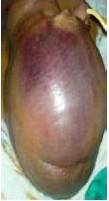
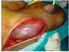
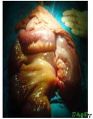
Giant hernias are rare in present times. They are present due to neglect (Patsas et al. 2010). The use of local anesthesia for hernioplasty techniques that encourage patients to undergo an operation shortly after diagnosis leads to rare occurrence of giant inguinal hernia today. The patient may remain asymptomatic or present with acute renal failure, perforation with concomitant peritonitis (Goonetille et al. 2010 and Gaedecke et al. 2013). In such giant hernia, ulceration, dermatitis, or candidiasis of scrotal skin could usually lead patients to seek medical care. There is a case report in which the incident of a gastric rupture in context with a giant left scrotal hernia has occurred (Walgenbach et al. 2001). Walking, sitting, simply lying down, and voiding may become extremely difficult for the patient. Penis could be buried, and only one testicle could be palpated. Usual content is gut, and sometimes the entire mesenteric small bowel and the entire colon may be lodged appendix, omentum, or bladder (Conda Sanchez et al. 2001 and Tahir et al. 2008). Rarely malrotated intestine could be present in a giant inguinal hernia (Lee 2012).
Due to rarity of condition, repair of giant inguinal hernia is always challenging, demanding to the surgeon, and stressful to the patient. Surgical management has to be tailored to the individual situation of the patient using all therapeutic options (Zippel et al. 2001). Loss of domain, recurrence and residual scrotal skin, and scrotal hematoma are the major problems encountered in management of such hernias (Coetzee 2011). A proper preoperative preparation for surgery in patients with giant hernias is desirable, especially involving respiratory status.
In elective repair, preoperative use of progressive pneumoperitoneum is effective in the treatment of large inguinal hernias (Piskin et al. 2010). Creation of pneumoperitoneum leads to optimal space for reduction of herniated contents into abdominal cavity and avoids abdominal morbidity in form of bowel resection, abdominal compartment, and extended abdominal wall reconstruction by the use of mesh (Vasiliadis et al. 2010). Laparoscopic component separation technique has been recommended to increase the capacity of the abdominal cavity to facilitate closure and reduce postoperative complications in patients who had loss of domain (Hamad et al. 2013).
El-Dessouki (2001) presented a technique for giant inguinal hernia in which hernia sac is pulled up to the abdomen and fashioned as a rotation flap to augment and close the peritoneum over the replaced contents; a giant polypropylene mesh is inserted in the preperitoneal space to cover the midline defect created and to buttress both inguinal regions. Zuvela et al. (2003) described the Rives technique (direct inguinal approach) in the treatment of large inguinoscrotal and recurrent hernias. Merret et al. (2009) advocated a technique for giant inguinal hernia involving the reduction of hernia; the repair of hernial orifices with Marlex mesh and the creation of a midline abdominal wall defect to increase the intra-abdominal capacity followed by covering this defect with Marlex mesh with a rotation flap of inguinoscrotal skin. Lichtenstein technique has also been advocated for repair of giant inguinal hernia (Bierca et al. 2013). A multistage operation for giant inguinal hernia with scrotal ulcer has been recommended where insufflation and prosthetic mesh are not available. In first stage, resection of ulcer and surrounding scrotal skin and partial reduction of hernia sac content are done. Partial reduction of hernia sac contents and resection of scrotal skins are done in stage two. In the last stage, bowel resection, ileocolic anastomosis, hernia repair, and resection of scrotal skin are done (Groen et al. 2011).
In an emergency, surgical treatment of giant abdominal hernias includes reduction of the hernia content and tension-free closure of the abdominal wall. Surgical treatment in complicated cases may require debulking the contents of the hernia sac by performing a right hemicolectomy and a small bowel resection and reconstruction of the abdominal wall using Marlex mesh and a tensor fasciae latae flap (Mehendal et al. 2000 and Goonetilleke et al. 2010). Inguinal incision aided by midline infraumbilical incision aids in the reduction of contents into abdominal cavity in giant inguinal hernia (Tahir et al. 2010).
Conclusion
Giant inguinal hernia presenting as intestinal obstruction is rare. Reduction of contents into abdominal cavity in giant inguinal hernia may be done by enlarging internal ring.
References
Bierca, J., Kosim, A., Małgorzata, K., Zmora, J. & Kultys, E. (2013). “Effectiveness of Lichtenstein Repairs in Planned Treatment of Giant Inguinal Hernia — Own Experience,” Videosurgery Miniinv, 8 (1) 36-42.
Publisher – Google Scholar
Coetzee, E., Price, C. & Boutall, A. (2011). “Simple Repair of a Giant Inguinoscrotal Hernia,” International Journal of Surgery Case Reports, 2(3)32-5
Publisher – Google Scholar
Conde Sánchez, J. M., Espinosa Olmedo, J., Salazar Murillo, R., Vega Toro, P., Amaya Gutiérrez, J., Alonso Flores, J. & García Pérez, M.(2001). “Giant Inguino-Scrotal Hernia of the Bladder. Clinical Case and Review of the Literature,”Actas Urologicas Espanolas, 25(4)315-9.
Publisher – Google Scholar
El-Dessouki, N. (2001). “Preperitoneal Mesh Hernioplasty in Giant Inguinoscrotal Hernias: A New Technique with Dual Benefit in Repair andAbdominal Rooming,” Hernia, 5(4)177-81.
Publisher – Google Scholar
Gaedcke, J., Schüler, P., Brinker, J., Quintel, M. & Ghadimi, M. (2013). “Emergency Repair of Giant Inguinoscrotal Hernia in a Septic Patient,” Journal of Gastrointestinal Surgery,17(4)837-9.
Publisher – Google Scholar
Goonetilleke, K. & McIlroy, B. (2010). “Giant Inguinoscrotal Hernia Presenting with Acute Renal Failure: A Case Report and Review ofLiterature,” Annals of The Royal College of Surgeons of England, 92(4) W21-3.
Publisher – Google Scholar
Groen, R., Sesay, S., Kushner, A. & Dumbuya, S. (2011). “Three-stage Repair of a Giant Inguinal Hernia in Sierra Leone: A Management Technique for Low-Resource Settings,” Journal of Surgical Case Reports. (12): 8
Publisher – Google Scholar
Hamad, A., Marimuthu, K., Mothe , B. & Hanafy, M. (2013). “Repair of Massive Inguinal Hernia with Loss of Abdominal Domain UsingLaparoscopic Component Separation Technique,” Journal of Surgical Case Reports. 3 (3pages)
Publisher – Google Scholar
King, J. N., Didlake, R. H. & Gray, R. E. (1986). “Giant Inguinal Hernia,” Southern Medical Journal, 79(2) 252-3.
Publisher – Google Scholar
Lee, S. E. (2012). “A Case of Giant Inguinal Hernia with Intestinal Malrotation,” International Journal of Surgery Case Reports, 3(11)563-4
Publisher – Google Scholar
Mehendal, F. V., Taams, K. O. & Kingsnorth, A. N. (2000). “Repair of a Giant Inguinoscrotal Hernia,” British Journal of Plastic Surgery, 53 (6)525-9.
Publisher – Google Scholar
Merrett, N. & Biankin, A. (2009). “Giant Inguinal Hernia Containing Right “Colon Repaired Using the Prolene Herniasystem,” ANZ Journal of Surgery, 79(1-2) 92-3.
Publisher – Google Scholar
Patsas, A., Tsiaousis, P., Papaziogas, B., Koutelidakis. I., Goula, C. & Atmatzidis, K. (2010). “Repair of a Giant Inguinoscrotal Hernia,” Hernia,14(3)305-7.
Publisher – Google Scholar
Piskin, T., Aydin, C., Barut, B., Dirican, A. & Kayaalp, C. (2010). “Preoperative Progressive Pneumoperitoneum for Giant Inguinal Hernias,” Annals of Saudi Medicine, 30(4)317-20.
Publisher – Google Scholar
Sarkarbi, W., Al. Agrawal, A. & Taffinder, N. (2005). “A Giant Inguinoscrotal Hernia: A Case Report and Review of the Literature,” Grand Rounds, 5:46-48
Publisher – Google Scholar
Tahir, M., Ahmed, F. U. & Seenu, V. (2008). “Giant Inguinoscrotal Hernia: Case Report and Management Principles,”International Journal of Surgery, 6(6)495-7.
Publisher – Google Scholar
Vasiliadis, K., Knaebel, H. P., Djakovic, N., Nyarangi-Dix, J., Schmidt. J. & Büchler, M. (2010). “Challenging Surgical Management of a Giant Inguinoscrotal Hernia: Report of a Case,” Surgery Today, 40(7)684-7.
Publisher – Google Scholar
Veihelmann, A., Ungeheuer, A. & Feussner, H. (2001). “Case Report: Emergency Surgery of a Giant Scrotal Hernia,”Zentralblatt für Chirurgie, 126(12) 1018-20.
Publisher – Google Scholar
Walgenbach, K.- J., Lauschke, H., Brünagel, G. & Hirner, A. (2001). “An Uncommon Form of Gastric Rupture in Giant Scrotal Hernia,” Zentralblatt für Chirurgie, 126(12)1015-7.
Publisher – Google Scholar
Zippel, R., Meyer, L., Kube, R. & Gastinger, I. (2001). “Elective Surgical Treatment of a Giant Scrotal Hernia,” Zentralblatt für Chirurgie, 126(12)1021-3.
Publisher – Google Scholar
Zuvela, M., Milićević, M., Lekić, N., Raznatović, Z., Palibrk, I., Bulajić, P., Petrović, M., Basarić, D. & Galun, D., (2003). “The Rives Technique (Direct Inguinal Approach) in Treatment of Large Inguino-Scrotal and Recurrent Hernias,” Acta Chirurgica Iugoslavica, 50(2)37-48.
Publisher – Google Scholar


