Tuberculosis or Sarcoïdosis. A Case Report

Raja Lihiou, Hajer Ben Brahim, Olfa Harzallah, Abir Aouam, Adnene Toumi, Chaouki Loussaïef and Mohamed Chakroun

Infectious disease department, Monastir, Tunisia

Academic Editor: Batool Sharifi - Mood

Cite this Article as:

Raja Lihiou, Hajer Ben Brahim, Olfa Harzallah, Abir Aouam, Adnene Toumi, Chaouki Loussaïef and Mohamed Chakroun (2013), "Tuberculosis or Sarcoïdosis. A Case Report," International Journal of Case Reports in Medicine, Vol. 2013 (2013), Article ID 772529, DOI: 10.5171/2013.772529

Copyright © 2013 Raja Lihiou, Hajer Ben Brahim, Olfa Harzallah, Abir Aouam, Adnene Toumi, Chaouki Loussaïef and Mohamed Chakroun. Distributed under Creative Commons CC-BY 3.0

Abstract

Distinguishing sarcoïdosis from pulmonary tuberculosis can be a great challenge to physicians, especially in Tunisia where there is high prevalence of tuberculosis. Both tuberculosis and sarcoïdosis are granulomatous diseases; however, necrosis is commun in tuberculosis and less common in sarcoïdosis. The presence of necrosis may lead to an excessive diagnosis of tuberculosis. We present the case of a 48 years old female with nasosinusal tuberculosis, based on granulomatous inflammation with little fields of necrosis. The patient was treated byanti-tubercular therapy for many months without healing. The diagnosis was revised when progressive pulmonary lesions were observed while the patient was on well conducted anti-tubercular treatment. Sarcoïdosis was finally retained with good response to corticosteroid therapy.   
Keywords: Tuberculosis, sarcoïdosis, granuloma.

Introduction

Systemic granulomatosis are frequently encountered in internal medicine. In these cases tuberculosis still the first diagnosis in tuberculosis endemic area like Tunisia. Other granulomatous diseases must be usually discussed, especially sarcoïdosis [Farheen et al., 2011]. In both clinical and histopathological features sarcoidosis is remarkably similar to tuberculosis and therefore can be difficult to distinguish. This case illustrates diagnostic difficulties between the two pathologies.

Observation

A 48 years old woman, with a history of right facial nerve palsy since the age of 12, had been followed in Otorhinolaryngology department (ORL) since 2003 for chronic nasal obstruction. In April 2008, nasal tuberculosis was diagnosed based on nasal mucosa biopsy showing granulomatous inflammation with necrosis. Both Ziehl-Neelsen stain and mycobacterium culture of the specimen were negative. In spite of the absence of impregnation symptoms, anti-tubercular therapy (ATT) was initiated in June 2008, because our country is endemic for tuberculosis, using the four routine antitubercular drugs: isoniazid (H), rifampicin (R), ethambutol (E) and pyrazinamid (Z). ATT was interrupted a few days later by the patient because of digestive intolerance.

In August 2008, ATT was represcribed associating HRZE during 4 months and then HR for 10 months, with little improvement. A nasal biopsy practiced, six months later, showed granulomatous inflammation with little necrotic fields. Then, the patient was transferred to the infectious diseases department for more exploration.

At admission, the patient has no symptom except nasal obstruction. The tuberculosis skin test was negative, sputum and gastric fluid examination and cultures for acid fast bacilli were negative. Serological tests for syphilis were negative. On chest X-ray, mediastinal enlargement was observed as well as interstitial lung disease in the lower part of the right lung (fig.1). Facial computed tomography scan showed: thickening of nasal cavity and right maxillary sinus mucosa (fig.2).

772529-fig-1

Fig.1. Mediastinal Enlargement ( ). Interstitial Disease with Irregular Opacity (    ). (Marsh 2011)

 
772529-fig-2

Fig.2. Thickening of Nasal Cavity and Right Maxillary Sinus Mucosa. (Marsh 2011)


Thoraco-abdominal computed tomography scan showed mediastinal enlarged lymph nodes and alveolar condensation with central excavation, inside of ground-glass pattern in the lower right lung field.

This presentation suggested progressive pulmonary tuberculosis occurring while the patient was on well conducted ATT. Subsequently, multi-resistant mycobacterium tuberculosis or atypical mycobacterium infection were suspected and a new ATT treatment protocol was prescribed associating Clarithromicin, Amikacin, Ethambutol, Rifampicin and Ofloxacin. In April 2011, non granulomatous anterior uveitis occurred and was controlled by corticoid eye drops and hypotonic treatment.

In November 2011, after seven months of ATT, nasal obstruction persisted and the patient was free of respiratory symptoms. Computed tomography showed: stable naso-sinusal findings, no modification in the size of mediastinal lymphadenomegalies, worsening in lung damage; irregular alveolar condensations of the right basis, peripheral nodules and distortion of pulmonary architecture with bronchectasies (fig.3).

772529-fig-3

Fig.3: Irregular Alveolar Condensations of the Right Basis. Distortion of Pulmonary Architecture with Bronchectasies (November 2011)


These findings were very suggestive of pulmonary sarcoïdosis isolated or added to tuberculosis.

In February 2012, the patient was hospitalized for exploration. She complained of moderate inflammatory joint pain, xerostomia, blurred vision and bilateral watering. She was free of fever, weight loss, dyspnea, cough, chest pain or hemopthysis.

Physical examination was normal aside from facial palsy.  Standard biological tests reveled: Hemoglobin rate 12.6 g/dl, white blood cell count 4300/mm3 with lymphopenia (800/mm3), platelet count 260.000/mm3, erythrocyte sedimentation rate 15 mm/h, C-reactive protein level 9 mg/l, hypergammaglobulinemia (15.5 g/l). Proteinuria was positive at 0.2 g/24h, urinalysis for bacteria was negative. Hepatic and renal tests were normal. Tuberculin skin test was anergic, mycobacterial tuberculosis bacilli were researched in sputum, gastric and bronchoalveolar lavage fluids with negative results. 

Chest X-ray showed mediastinal enlargement with fibro-indurative changes in the area of the lower part of the right lung with traction and a focus of «round glass» pattern.

Since sarcoïdosis was suspected, oriented tests were realized; calcium levels in plasma and urine were normal varying from 2.24 to 2.34 mmol/l and 0.06 to 0.07 mmol/kg/24h respectively. Angiotensin-converting enzyme (ECA) was elevated at 165 U/l (12-68 U/l). Pulmonary function test was normal. In broncho-alveolar lavage (BAL), macrophages were predominant. No histological lesion was noted on salivary glands biopsy.

Ophthalmologic examination noted: ocular hypertension, sequels of bilateral anterior uveitis with large basis irido-cornean synechia. According to ophthalmologists, these findings suggest tuberculosis or sarcoïdosis. Visual fields and color vision were normal.

AAN and ANCA search was negative. Since sarcoïdosis was suspected, corticosteroid therapy was initiated in March 2012, justified by naso-sinusal localization. On the other hand, thoracic lesions since asymptomatic with no functional impact, did not justify corticotherapy. The patient was treated by prednisolone 0.5 mg/kg body weight/day, associated to ATT which was stopped one month later. Improvement in nasal obstruction was remarkable within 2 weeks. The patient became totally asymptomatic six months later. After 12 months patient still asymptomatic under corticosteroid therapy (prednisone, 7.5 mg/day). Cranial and thoraco-abdominal computed tomography scan showed an important improvement in pulmonary and sinusal lesions (fig.4).

772529-fig-4

Fig.4: Improvement of Pulmonary Lesion with Corticosteroid Therapy

Discussion

Due to the marked clinico-radiological and histological similarity between tuberculosis and sarcoïdosis, and the high prevalence of tuberculosis in our country, confusion may occur and the patients may receive unuseful repeated courses of anti-tubercular therapy [Farheen et al., 2011].

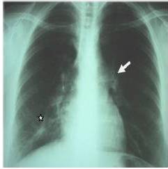
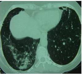
 Sarcoïdosis is a multisystem disorder of unknown cause, characterized by non specific constitutional symptoms such as fever, weakness and weight loss. Commonly, the disease involves mediastinal lymph nodes and lungs (90% of patients) but any other systems may be involved [Goksel et al., 2011].The chest X-ray is abnormal in 85 to 95% of cases. It commonly shows hilar and mediastinal lymphadenopathy. Other characteristic findings include interstitial lung disease, retractions and irregular opacities. Computed-tomography scan of the thorax shows thickening of bronchovascular bundles and perilymphatic distribution of nodules which represent the sarcoïd granuloma [Valeyre et al., 2005]. At the stage of fibrosis: bronchial distortion, hilar retraction, diaphragmatic ascension and bronchectasies are noted. Perihilar masses rarely evolve toward the excavation. Aspects of ray honey, ground glass opacities and bubbles are less frequently observed [Valeyre et al., 2005]. The tuberculin anergy is present in 75 % of cases. Generally there is no biological inflammatory syndrome. The hypergammaglobulinaemia is common. Lymphopenia, hypercalciuria, more rarely hypercalcemia may contribute to the diagnosis. The diagnostic value of ECA level is low [Pavic et al., 2008]. At BAL, lymphocytes rate over than 30 % (normal = 10 — 15 %) increases the probability of sarcoïdosis.

Histopathologically, sarcoïdosis is characterized by the presence of non-caseating granuloma. Rarely, non-caseating necrosis may be noted on sarcoïdosis granuloma, in this case little and fibrinoid [Pavic et al., 2008].

At initial presentation the epidemiological and clinical features, presence of necrosis in side granuloma could very well be matched with the diagnosis of tuberculosis. Presence of cavity formation, common in TBC and uncommon in sarcoïdosis, strengthens this hypothesis. Repeated courses of ATT with whole duration of 25 months were instituted while lung damage continues to progress with new radiological findings more suggestive of sarcoïdosis such as peripheral nodules and distortion of pulmonary architecture with bronchectasies.

After oriented check-up, it has been clear that the patient had many features of sarcoïdosis, with usually negative reaction to tuberculin and no evidence of active tuberculosis. Mycobacterium tuberculosis was never isolated. Finally the diagnosis was redressed and the favorable evolution with corticosteroid therapy confirms this hypothesis.

Recently, adding to the diagnostic difficulties, there are also some reports of coexistence of sarcoïdosis and tuberculosis in the same patients [Abdurrahman et al., 2006]. Tuberculosis has been described as both preceding and coexisting with sarcoïdosis, and as an opportunistic infection in patients with sarcoïdosis who are on corticosteroids, which depress the cell-mediated immunity. Only a few cases of TBC in patients with sarcoïdosis have been reported in the world literature, and all of them had a course of corticosteroids or were immunosuppressed. Supriya and al (2010) relate a case of a patient, considered the unique in the literature, with sarcoïdosis, who had never taken corticosteroids and who develops tuberculosis. Fite and al (2006) demonstrated that M. tuberculosis DNA was present in 9/23 sarcoïdosis biopsies (39 %), in 1/23 control patients (4%) (p = 0.01), and in all tissue samples from 12 control patients with tuberculosis. These results suggest a causality link between M. tuberculosis infection and sarcoïdosis in some cases.

It is possible that a coincidental reactivation of tuberculosis with acute sarcoïdosis occurred. However, in some cases there may be an etiological relationship between sarcoïdosis and mycobacterial infection, and it is possible that clinical manifestations of sarcoïdosis may have been a response to infection with M. tuberculosis [Uddenfeldt et al., 1982].

Conclusion

Clinical and radiological features of tuberculosis and sarcoïdosis are quite similar. If we have no bacteriological nor histological proof for tuberculosis or in case of no improvement after ATT, diagnosis may be quickly reconsidered.

References

Abdurrahman, T., Ortenca, R., Arzu, S., Arzu, T., Ibrahım, H. & Lutfi, Ç.  (2006). “Concurrent Presentations of the Sarcoidosis, Tuberculosis and Lymphoma in a Single Patient,” Respiratory Medicine, 100, 951—953.
PublisherGoogle Scholar

Badar, F., Azfar, S. F.,Ahmad, I.,Yasmeen, S. & Kirmani, S. (2011). “Diagnostic Difficulties in Differentiating Sarcoidosis from Tuberculosis,” Oman Medical Journa, 26 (3), 210-211.
PublisherGoogle Scholar

Fité, E., Fernández-Figueras, M. T., Prats, R., Vaquero, M. & Morerea, J. (2006). “High Prevalence of Mycobacterium Tuberculosis DNA in Biopsies from Sarcoidosis Patients from Catalonia, Spain,” Respiration, 73, 20—26.
PublisherGoogle Scholar

Kiter, G.,Müsellim, B.,Cetinkaya, E.,Türker, H., et al. (2011). “Clinical Presentations and Diagnostic Work-up in Sarcoidosis: A Series of Turkish Cases (Clinics and Diagnosis of Sarcoidosis,” Tüberküloz ve Toraks Dergisi, 59 (3), 248-258.
PublisherGoogle Scholar

Pavic, M., Le Pape, E., Debourdeau, P., Rabar, D., Crevon, L. & Colle, B. (2008). “Granulomatoses Systémiques Pseudosarcoïdosiques D’étiologie Déterminée et non Tuberculeuse. Etude de 67 Observations,” La Revue de médecine interne, 29, 5—14. 
PublisherGoogle Scholar

Sarkar, S., Saha, K. & Das, C. S. (2010). “Isolated Tuberculous Liver Abscess in a Patient With Asymptomatic Stage I Sarcoidosis,” Respiratory Care, 55 (12), 1751—1753. 
PublisherGoogle Scholar

Uddenfeldt, M., Stjemberg, N. & Lundgren, R. (1982). “Sarcoidosis or Tuberculosis — A Case Report,” Tubercle, 63, 221-223. 
PublisherGoogle Scholar

Valeyre, D., Nunes, H., Duperron, F., Soler, P., Kambouchner, M. & Brauner, M. (2005). “Sarcoïdose,” EMC-Pneumologie2, 147—164.
PublisherGoogle Scholar 

Shares