Concurrent Spontaneous Umbilical and Abdominal Wall Endometriosis Retracting the Surrounding Cutis

Arianna Pacchiarotti1, Giusi Natalia Milazzo1, Vincenzo Gentile1, Paola Frati2, Chiara Assorgi1, Donatella Caserta1 and Massimo Moscarini1

1Department of gynecological obstetric and urological sciences, Faculty of Medicine and Dentistry, ‘Sapienza’ University of Rome, Sant’Andrea Hospital, Rome, Italy

2Department of anatomical, histological, forensic medicine and musculoskeletal sciences, “Sapienza” University of Rome, Sant’Andrea Hospital, Rome, Italy

Academic Editor: Warren G. Foster

Cite this Article as:

Arianna Pacchiarotti, Giusi Natalia Milazzo, Vincenzo Gentile, Paola Frati, Chiara Assorgi, Donatella Caserta and Massimo Moscarini (2013), "Concurrent Spontaneous Umbilical and Abdominal Wall Endometriosis Retracting the Surrounding Cutis," International Journal of Case Reports in Medicine, Vol. 2013 (2013), Article ID 994933, DOI: 10.5171/2013.994933

Copyright © 2013 Arianna Pacchiarotti, Giusi Natalia Milazzo, Vincenzo Gentile, Paola Frati, Chiara Assorgi, Donatella Caserta and Massimo Moscarini. Distributed under Creative Commons CC-BY 3.0

Abstract

Endometriosis consists of extra-uterine functional endometrial tissue. It is usually located in the pelvis, but it can also be found in other sites. Cutaneous endometriosis is a rare condition and it develops in the most of cases above gynecological or obstetric scars, although it may also appear spontaneously. We present a 39-year-old woman with umbilical and abdominal dermal nodules retracting the surrounding cutis as a clinically characteristic form of spontaneous cutaneous endometriosis. The patient had no signs and symptoms of pelvic endometriosis. The histopathological and immunohistochemical examinations confirmed the clinical diagnosis.
Keywords: Umbilical endometriosis, cutaneous endometriosis, spontaneous extrapelvic endometriosis.

Introduction

Endometriosis is a common gynecological disease, defined as the presence of functional endometrial tissue outside the uterine cavity (Burney, 2012). Many etiological hypothesis have followed one another over the years, from the first theory of retrograde menstruation proposed by Sampson in 1927 (Sampson, 1927) to the recent theories involving a stem cell origin (Pacchiarotti, 2011). Endometriosis generally involves the ovary and the pelvic peritoneum, but in 9-12% of cases it reaches extragonadal sites (Donnez, 2004), such as bowel, bladder, lungs (Kodandapani, 2011) and nerves (Pacchiarotti, 2013). Cutaneous endometriosis represents less than 5.5% of all cases of endometriosis (Din, 2013) and umbilical endometriosis 0.5-1% of extragonadal endometriosis (Kodandapani, 2011). Cutaneous endometriosis is mostly associated with surgical scars (60% of cases), less frequently it occurs spontaneously (Singh, 2012). Clinical diagnosis can be difficult due to its rarity and the lack of pathognomonic signs except for its changes during the menstrual cycle. Correct pre-operative diagnosis is attained in approximately 25% of cases (Din, 2013).

Case Report

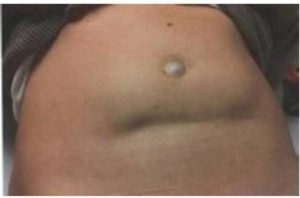
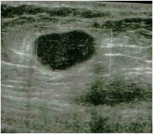
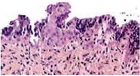
A 39-year-old woman reported a blue swelling at the navel and two subcutaneous nodules with bluish transparency on the suprapubic line. She described that swellings changed during menstruation. Umbilical nodule showed a diameter of about 1.5 cm; it was soft, but not reducible, and dark. Both lumps on the pubic line appeared rather deeper, irreducible, in a bluish transparency and they formed an area of skin retraction which joined the two nodules in an horizontal line (Fig. 1). The patient had no previous abdomen surgery neither typical endometriosis symptoms, such as dysmenorrhea or dyspareunia. Abdominal wall ultrasound (US) of the nodules showed well defined anechoic areas (Fig. 2).  Suspecting endometriosis, we performed US transvaginal examination, which showed no signs of the disease. Because of pain due to the umbilical lesion, we decided to remove it surgically.  The umbilical nodule was sent for histological examination, which confirmed the presence of a dermal endometriosis (Fig. 3) and immunohistochemical staining for estrogen and progesterone receptors was positive.

Discussion

This is a rare case of concomitant spontaneous umbilical and abdominal wall endometriosis. The most common site of spontaneous endometriosis is the umbilicus, followed by the inguinal area and the abdominal wall (Kyamidis, 2011). Cutaneous endometriosis could be suspected in women of reproductive age when there is a palpable abdominal bluish nodule, characterized by cyclic pain and swelling. In our case also abdominal skin retraction, due to nodules’ cyclical bleedings and subsequent fibrosis, may guide the diagnosis. Non specific chronic suppuration (Din, 2013) and bleeding (Bagade, 2009) may be another clinical manifestation of umbilical endometriosis. A history of pelvic pain and sterility may corroborate the hypothesis of endometriosis (Kyamidis, 2011).The localization on a surgical scar may be suspicious of a secondary endometriosis (Fernàndez Vozmediano, 2010). Abdominal wall US is useful to demonstrate the cystic nature of the lesion and the MRI and CT can help to assess the extent of the disease and differential diagnosis with hernias (Din, 2013). The differential diagnosis should include lipoma, dermoid cyst, haemangioma, keloid, hernia, abscess, pyogenic or foreign granuloma, embryological rests, irreducible hernia, inclusion cyst, metastatic tumors from intra-abdominal malignancy and melanoma (Din, 2013; Kyamidis, 2011; Singh, 2012). Fine needle aspiration cytology may be conducted (Fernandes, 2011) even if histology is the best diagnostic tool, supported by  immunohistochemical analysis of estrogen and progesterone receptors when the excessive fibrosis hides field (Kyamidis, 2011).The complete excision of the lesion, under local or loco-regional anesthesia, is the treatment of choice and usually curative. Hormonal therapies are insufficient as sole treatments (Kyamidis, 2011). These latter may be an alternative treatment for little endometriomas or may be used before surgery to reduce the size of lesions (Kyamidis, 2011). Local recurrence after adequate surgical excision is rare. Malignant transformation has been described (Chene, 2007). Recognizing cutaneous endometriosis is very important for a prompt endometriosis’ diagnosis, which is generally gained with a 6.7-year delay (Burney, 2012). As endometriosis is a progressive disease, delaying diagnosis and treatment would increase the risk of severe pain, distortion of the pelvic anatomy and sterility.

994933-fig-1

Figure 1:  Clinical Appearance of the Lesions before the Surgical Treatment

994933-fig-2

Figure 2: US Image of One Dermal Nodule of the Lower Abdominal Wall (1.52×0.92 Mm)

994933-fig-3


Figure 3: Histological Examination 

References

Bagade, P. V. & Guirguis, M. M. (2009). “Menstruating from the Umbilicus as a Rare Case of Primary Umbilical Endometriosis: A Case Report,” Journal of Medical Case Reports, 3: 9326.
PublisherGoogle Scholar

Burney, R. O. & Giudice, L. C. (2012). “Pathogenesis and Pathophysiology of Endometriosis,” Fertility and Sterility, 98 (3): 54-9.
PublisherGoogle Scholar

Chene, G., Darcha, C., Dechelotte, P., Mage, G. & Canis, M. (2007). “Malignant Degeneration of Perineal Endometriosis in Episiotomy Scar, Case Report and Review of the Literature,” International Journal of Gynecological Cancer, 175 (3) : 709-14.
PublisherGoogle Scholar

Din, A. H., Verjee, L. S. & Griffiths, M. A. (2012). “Cutaneous Endometriosis: A Plastic Surgery Perspective,” Journal of Plastic, Reconstructive & Aesthetic Surgery, 66 (1) : 129-30
PublisherGoogle Scholar

Donnez, J. & Van Langendonckt, A. (2004). “Typical and Subtle Atypical Presentations of Endometriosis,” Current Opinion in Obstetrics and Gynecology, 16(5):431-437.
PublisherGoogle Scholar

Fernandes, H., Marla, N. J., Pailoor, K. & Kini, R. (2011). “Primary Umbilical Endometriosis – Diagnosis by Fine Needle Aspiration,” Journal of Cytology, 28 (4) : 214-6.
PublisherGoogle Scholar

Fernández Vozmediano, J. M., Armario Hita, J. C. & Cuevas Santos, J. (2010). “Cutaneous Endometriosis,” International Journal of Dermatology, 49 (1) : 1410-2.
PublisherGoogle Scholar

Kodandapani, S., Pai, M. V. & Mathew, M. (2011). “Umbilical Laparoscopic Scar Endometriosis,” Journal of Human Reproductive Sciences, 4 (3) : 150—152.
PublisherGoogle Scholar

Kyamidis, K., Lora, V. & Kanitakis, J. (2011). “Spontaneous Cutaneous Umbilical Endometriosis: Report of a New Case with Immunohistochemical Study and Literature Review,” Dermatology Online Journal, 17 (7) : 5.
PublisherGoogle Scholar

Pacchiarotti, A., Caserta, D., Sbracia, M. & Moscarini, M. (2011). “Expression of Oct-4 and c-kit Antigens in Endometriosis,” Fertility and Sterility, 95 (3) : 1171-3.
PublisherGoogle Scholar

Pacchiarotti, A., Milazzo, G. N., Biasiotta, A., Truini, A., Antonini, G., Frati, P., Gentile, V., Caserta, D. & Moscarini M. (2013). “Pain in the Upper Anterior-Lateral Part of the Thigh in Women Affected by Endometriosis: Study of Sensitive Neuropathy,” Fertility and Sterility [epub ahead of print]
PublisherGoogle Scholar

Sampson, J. A. (1927). ‘Peritoneal Endometriosis Due to Menstrual Dissemination of Endometrial Tissue into the Peritoneal Cavity,’ American Journal of Obstetrics and Gynecology, 14:442—69.

Singh, A. (2012). “Umbilical Endometriosis Mimicking as Papilloma to General Surgeons: A Case Report,” Australasian Medical Journal, 5 (5) : 272-4.
PublisherGoogle Scholar

Shares