Neuropathological Study of Acute Myelopathy and Encephalopathy Associated to HTLV-I

Luis Cartier1, Carmen Vergara1, Eugenio Ramírez2 and María Antonieta Valenzuela3

1Departamento de Ciencias Neurológicas, Facultad de Medicina, Universidad de Chile, Chile

2Departamento de Virología, Instituto de Salud Pública de  Chile, Chile

3Departamento de Bioquímica y Biología Molecular, Facultad de Ciencias Químicas y, Farmacéutica, Universidad de Chile, Chile

Academic Editor: Rui Fernando M. Silva

Cite this Article as:

Luis Cartier, Carmen Vergara, Eugenio Ramírez and María Antonieta Valenzuela (2012), "Neuropathological Study of Acute Myelopathy and Encephalopathy Associated to HTLV-I," International Journal of Case Reports in Medicine, Vol. 2012 (2012), Article ID 996020, DOI: 10.5171/2012.996020

Copyright © 2012 Luis Cartier, Carmen Vergara, Eugenio Ramírez and María Antonieta Valenzuela. Distributed under Creative Commons CC-BY 3.0

Abstract

Background a chronic and progressive parapares is without remission silent on MRI is the ordinary neurological presentation of HTLV-I, expression of a central axonopathy product of axoplasmic transport alterations. Infrequently acute forms like “T2 hyperintense acute myelopathy on MRI” have been reported. The Purpose of this presentation will be to describe the histopathological expression of this acute form and look for their pathogenesis. Patient and method: A 60 years old woman carrier of refractory anaemia and subjected to multiple transfusions, positive for HTLV-I.  She developed a severe cognitive impairment and paraplegia set up in four weeks. MRI showed T2 hyper intense lesions in white matter of brain hemispheres and in cervico-thoraxic segments of the spinal cord. She died suddenly by myocardial infarction. The neuropathological studies showed white matter necrosis insymmetricfrontal areas, conservation of cortical structures and U fibbers; necrosis in bothlateral tracts of the spinal cord (T2- T4) without gray substance damage; the microvascular walls of these areas immuno-stained with anti-Tax, expressed Tax- protein suggesting HTLV-I infection.Conclusions: This necrotizing leukopathy in absence of a primary inflammation would be the expression of HTLV-I endothelial cells infection in specific white matter areas, changing of the blood-brain barrier permeability.  
Keywords: Endothelial HTLV-I infection. HTLV-I acute encephalomyelopathy. Necrotizing-encephalomyelopathy.

Introduction

The HTLV-I associated myelopathy (HAM) or tropical spastic paraparesis (TSP) was primarily communicated by Gessain et al (1985), from Martinique island patients.

This slowly and progressive spastic paraparesis with neurogenic bladder without remissions evolve with normal cerebrospinal fluid; few cases show mild lymphocytic pleocitosis; frequently blood count show leukemoid lymphocytes (Osame 1987); spinal cordis silent on MRI, however some cases show thoracic spinal cord atrophy after years (Yukitake 2008); most of the patients have somatosensory evoked potentials alterations (Castillo1999). This chronic paraparesis is a degenerative disease (Cartier 1997) expression of a central axonopathy, defined by axonal torpedoes, deposits of amyloid precursor protein (APP) and loss of the axons, (Cartier 2007). The virus may also engage the cerebral white matter, and has been reported HAM/TSP with progressive cognitive impairment and dementia (Ogata 1993).

Frequently these patients present associated diseases, the half of them shows sicca syndrome (Cartier 1995/a); almost 10%of patients may develop some type of lymphoma, often smouldering lymphoma (Cartier 1995/b); less common is the chronic hepatitis, or liver cirrhosis associated to HTLV-I (Ijichi 1993); the osteoporosis is a frequent and non apparent complication (Schachster 2003); others associated diseases, like uveitisor polymyositis have been described (Nakagawa 1995).

Definitively the ”T2 hyper intense acute myelopathy on MRI” is uncommon manifestation of the CNS HTLV-I infection (Sakudo 1995, Watanabe 2001), the clinical evolution of these acute or sub acute forms would be suggesting different pathogenetic expression that the chronic form.

We are showing the neuropathological study of simultaneous HTLV-I acute myelopathy and encephalopathy.

Case Report

The studied patient is a sixty years old woman, carrying of a refractory anemia, subject to numerous blood or red corpuscles transfusion, in the last three years. Four weeks before her admissions he began with a cognitive impairment, expressed as a mnemonic failure and intellectual decay, that added gait difficulties a week before her admission. She entered with a paraplegic recently installed, and a demential syndrome. The patient was disoriented, had difficulty to understand and execute simple commands, memory was not evaluable; she had a paraplegia with bilateral Babinski, sensory level at third thoracic and neurogenic bladder. The brain MRI on T2 showed hiperintensity in white matter of both cerebral hemispheres, with greater frontal expression. (Fig.1)The spinal cord MRI on T2 shows hiperintensity and thickening (C7 toT4) (Fig. 2).  The EEG expresses theta activity and amplitude of 100mv. The general examination identified hepatomegaly and splenomegaly. The hematocrit was of 20%, hemoglobin 7.1 gr., leukocyte 3620, ferritin 4280ug, total bilirubin 1.91 gr. /dl, 63% prothrombinemia. Anti Nuclear antibody (ANA) and Antineutrophil cytoplasmic antibodies (ANCA) were negatives. We ruled out syphilis, virus Herpes, JC, and HIV. The HTLV-I was positively identified in PBMCs by real-time PCR and flow cytometry respectively. Normal cerebrospinal fluid was positive to HTLV-I in western blot. Abdominal scan and ECO showed hyperdensity of liver. Myelodysplastic myelogram, define a refractory anemia with ringed sideroblasts. Schirmer’s test pointed out hipolacrimia (6mm OD and 7mm OI). Immunohistochemistry of minor salivary gland biopsy proved the presence of Tax protein in glandular acini and underlying lymphocytes. Patient died suddenly of a heart attack in the third week of hospitalization. Pathology: The immediate cause of death was acute myocardial infarction. We also found an old sub-endocardial infarction and ventricular hypertrophy. Also it was defined a lymphoma present in lymph nodes. The liver showed intracellular hemosiderin deposits, suggesting a secondary hemochromatosis. Neuropathology: The brain weighted 1355 grams, had normal external appearance, and was fixed in formalin10% showing cortexpreservation and white matter changes especially in both frontal areas. (Fig.3) Basal ganglia, sub-thalamic region, cerebellum and brainstem were preserved. The spinal cord was thickened and soft in high dorsal segments. Microscopic study was performed with hematoxylin eosin (HE), Luxol Fast-blue (LFB), Van Gieson (VG) and GFAP. Immunohistochemistry study for neurofilaments, we used (Monoclonal Mouse Anti-Human Neurofilament Protein Clone 2F11/ Code-Nr. M 0762 DAKO (1:100)); for Tax protein, mab0022 HTLV-1 Tax, COVALAB (1:50)); for CD45M0833Dako (1:50)); CD4 SP35 CELL MARQUE (1:50)); CD8 Clone C8/144B DAKO (1:20)); Ubiquitin NCL UBIQ NOVOCASTRA (1:50));Sumo ( Anti-Sumo 1 antibody (ab11291) Rabbit polyclonal to Sumo 1 Abcam (1 :1000)); Semaphorin ( Anti-Semaphorin 4D antibody Rabbit polyclonal  Abcam (ab39710) (1:200)).

996020-fig-1

Fig. 1: Magnetic Resonance: A) White Matter Damage Predominant in Frontal Areas (T1). — B) Hyperintensity of White Matter in Both Cerebral Hemispheres. (FLAIR)

996020-fig-2

Fig. 2: Spinal Cord Magnetic Resonance: Fusiform Hyperintensity from C7 to T4 and Expanding of the Parenchyma of Spinal Cord in that Area. (T 2)

996020-fig-3

Fig. 3: -Macroscopic Appearance: The White Matter of Frontal Areas Shows Necrotic Lesions in Both Hemispheres.

 
Both cerebral hemispheres show white matter necrosis in the oval center prevailing in frontal areas; the cortical structures and the U fibbers are preserved. (Fig. 4) Spinal cordnecrosis was found in lateral tracts of thoracic segments (T2 – T4), without compromise of gray substance (Fig. 5) Cerebral and spinal cord lesions give the appearance of ischemic damage showing granular bodies, gemistocytic astrocytosis and vascular thickening in the central part of lesions (Fig.6), laterally spongiosis and myelin pallor were seen. Wallerian degeneration of affected lateral columns was expressed lower dorsal and lumbar. In damaged areas immunore activity with anti-Tax was expressed on the vascular wall and detected some infected lymphocytes. (Fig. 7) Anti-neurofilaments immunohistochemistry shows axonal swellings and characteristic fragmentation of them. Anti-ubiquitin reaction was found in perivascular areas; also related with some axons (not show). Semaphorin stained oligodendrocytes close to the lesion, and gemistocytic glia (not show). Necrotic damage of cerebral hemispheres and in both lateral tracts of spinal cordexpressesa systemic compromise of the white matter.

996020-fig-4

Fig. 4: Histological Study: Damages in Frontal White Matter, Necrosis Encircled by Spongiotic and Demyelinated Areas, which Take the Appearance of Ischemic Injury. The U Fibbers and the Cortex are Preserved (Haematoxylin and LFB x 40).

996020-fig-5

Fig. 5: Histological Study: The First Thoracic Segments of Spinal Cord, Showing Focal Necrosis of the Lateral Cord and Preservation of Gray Matter and Posterior Columns. (LFB and Haematoxylin x 40

996020-fig-6
Fig. 6: Histological Study: Shows Necrotic Area of the White Matter in the Absence of Primary Inflammation, Granular Bodies, Thickened Vessels and Reactive Gliosis are Seen (HE x 150).

996020-fig-7

Fig.7: Histological Study: Shows Tax Protein Expression in the Vascular Wall of the Lesional Areas.-A) Anti-Tax x200.-B) Anti-Tax and Haematoxylin x 200.

 
Comment

Simultaneous acute encephalopathy and myelopathy hyperintense in T2 on MRI is presented. The hyperintensity in T2 on MRI seems a characteristic finding in acute cases. Sakudo et al (1999) were the firsts to identify these clinical forms of HTLV-I paraparesis with abnormalities on MRI. Watanabe et al (2001) reported one case with MRI abnormalities and rapid progression of the paraparesis. Two acute cases and MRI alterations were reported by Tajima et al. (2003). The neuro imaging analysis of 38 patients with HAM / TSP, performed by Yukitake et al (2008) showed 22 cases without spinal cord alterations, 13 patients exhibited atrophy of the thoracic spinal cord and only 3 cases show T2 hyperintense lesions, defined by them like “malignant” cases.

Necrosis of specific areas in the white matter as in cerebral hemispheres, as in lateral tracts of the spinal cord are suggesting different pathogenesis for these acute or subacute forms respect to chronic HAM/TSP.

To understand this different and particular expression is necessary to know the retrovirus effect over endothelial cells of CNS vessels. The infection of endothelial and glial cells of the blood-brain barrier stops the genesis of specific proteins and then the blood – brain barriers alter their specific permeability in those affected areas. In HTLV-I patients this infection is facilitated because the receptors for HTLV-I are expressed in endothelial cells of the CNS vessels. (Afonso 2008 and 2007)

The symmetric lesions of this studied-case show evidence that myelopathy and encephalopathy would be a consequence of microvascular compromise in “privileged” areas in absence of primary inflammatory lesions. This form of white matter necrosis resembles those observed in Devic disease caused by aquaporin dysfunction on specifics areas of the spinal cord and others, where they produced changes in the BBB, and necrotic lesions (Jairus 2008).

In this acute form is interesting to remarque a distalwallerian degeneration of the injured tracts in the spinal cord, fact not observed in the chronic form. (Cartier 1997).

The HTLV-I infected patients seems to have a latent double pathogenic mechanism, « vascular » by changes in BBB and  « toxic »by  sustained increase of HTLV-I Tax protein over axoplasmic transport (Alberti 2011). The clinical and pathological expression will be subordinate which of them prevail, dependant the virus activity and the host condition.

More studies will be necessaries to confirm this suggestive hypothesis, which could explain the acute neurological compromise of HTLV-I infections.

References

Afonso, P. V., Ozden, S., Cumont, M.- C., Seilhean, D., Cartier, L., Payam, R., Mason, S., Lambert, S., Huerre, M., Gessain, A. et al. (2008). “Alteration of Blood-Brain Barrier Integrity by Retroviral Infection,” PloS Pathogens, 4: 1-11.
PublisherGoogle Scholar

Afonso, P. V., Ozden, S., Prevost, M. C., Schmitt, C., Seilhean, D., Weksler, B., Couraud, P. O., Gessain, A., Romero, I. A. & Ceccaldi, P. E. (2007). “Human Blood-Brain Barrier Disruption by Retroviral-Infected Lymphocytes: Role of Myosin Light Chain Kinase in Endothelial Tight-Junction Disorganization,” The Journal of Immunology, 179; 2576-2583.
PublisherGoogle Scholar

Alberti, C., Cartier, L., Valenzuela, M., Puente, J., Tanaka, Y. & Ramirez, E. (2011). “Molecular and Clinical Effects of Betamethasone in Human T-cell Lymphotropic Virus Type-I-Associated Myelopathy/Tropical Spastic Paraparesis Patients,” Journal of Medical Virology 83:1641-9.
PublisherGoogle Scholar

Cartier, L., Castillo, J. L., Cabrera, M. E., Araya, F., Matutes, E., Ford, A. M. & Greaves, M. F. (1995/b). “HTLV-I Positive Progressive Spastic Paraparesis (TSP) Associated with a Lymphoid Disorder in Three Chilean Patients,” Leukemia and Lymphoma, 17: 459-464.
PublisherGoogle Scholar

Cartier, L., Castillo, J. L., Cea, J. G. & Villagra, R. (1995/a). ‘Chronic Dacrosyalpadenitis in HTLV-l Associated Myelopathy,’ Journal of Neurology, Neurosurgery & Psychiatry, 58: 244-246.

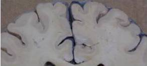
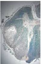
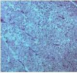
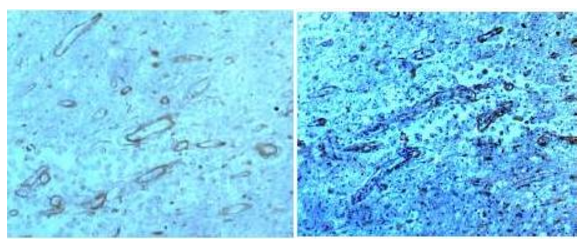
Cartier, L., Cea, J. G., Vergara, C., Araya, F. & Born, P. (1997). “Clinical and Neuropathological Study of Six Patients with Spastic Paraparesis Associated with HTLV-I an Axomyelinic Degeneration of the Centralnervous System,” Journal of  Neuropathology & Experimental Neurology, 56:403—413.
PublisherGoogle Scholar

Cartier, L.,Vergara, C. & Valenzuela, M. A. (2007).  “Inmunohistoquímica de Los Cambios Degenerativos del Sistema Nervioso Central en Paraparesias Espásticas Asociadas al Virus Linfotrópico Humano T Tipo I (HTLV-I),” Revista Médica de Chile,135: 1139-1146.
PublisherGoogle Scholar

Castillo, J. L., Cea, J. G., Verdugo, R. J. & Cartier, L. (1999). “Sensory Dysfunction in HTLV-I Associated Myelopathy/Tropical Spastic Paraparesis (HAM/TSP): A Comprehensive Neurophysiologic Study,” European Neurology42:17-22.
PublisherGoogle Scholar

Gessain, A., Barin, F., Vernant, J. C., Gout, O., Calender, A. & De The G. (1985). ‘Antibodies to Human T-lymphotropic Virus in Patients with Tropical Spastic Paraparesis,’ Lancet, U:407-410.

Ijichi, T., Miyata, K., Mori, S., Nakajima, K., Okanoue,T. & Tsuchihashi, Y. (1993). “Asymptomatic Primary Biliary Cirrhosis in HTLV-l Associated Myelopathy,” The American Journal of Gastroenterology, 88; 2107-2109
Publisher

Jairus, S., Paul, F., Franciotta, D., Waters, P., Zipp, F., Hohlfeld, R., Vincent, A. & Wildemann, B. (2008). “Mechanisms of Disease: Aquaporin-4 Antibodies in Neuromyelitisóptica,” Nature Clinical Practice Neurology, 4: 202-214
Publisher

Nakagawa, M., Izumo, S., Ijichi, S., Kubota, H., Arimura, K., Kawabata, M.& Osame, M. (1995). “HTLV-I-Associated Myelopathy: Analysis of 213 Patients Based on Clinical Features and Laboratory Findings,” Journal of Neurovirology, 1:50—61.
PublisherGoogle Scholar

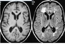
Ogata, A., Nagashima, K., Tashiro, K., Miyakawa, A. & Mikuni, C. (1993). “MRI Pathological Correlate of Brain Lesions in a Necropsy Case of HTLV-I Associated Myelopathy,” Journal of Neurology, Neurosurgery & Psychiatry, 56:194-196.
PublisherGoogle Scholar

Osame, M., Matsumoto, M., Usuku, K., Izumo, S., Ijichi, N., Amitani, H., Tara, M. & Igata, A. (1987). “Chronic Progressive Myelopathy Associated with Elevated Antibodies to Human T-Lymphotropic Virus Type I and Adult T-cell Leukemia-Like Cells,” Annals of Neurology 21: 117-122
PublisherGoogle Scholar

Schachter, D., Cartier, L. & Borzutzky, A. (2003). “Osteoporosis in HTLV-I Associated Myelopathy/Tropical Spastic Paraparesis (TSP/HAM),” Bone, 33: 192-96
PublisherGoogle Scholar

Shakudo, M., Inoue, Y. & Tsutada, T. (1999). “HTLV-I-Associated Myelopathy: Acute Progression and Atypical MRI Findings,” American Journal of Neuroradiology, 20: 1417-1421.
Publisher

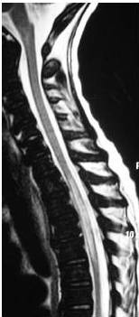
Tajima, Y., Kishimoto, R., Sudoh, K., Matsumoto, A., Miyazaki, Y., Kikuchi, S. et al. (2003). “Spinal Magnetic Resonance Image Alterations in Human T Lymphotropic Virus Type I-Associated Myelopathy Patients before and after Immunomodulating Treatments,” Journal of Neurology, 250: 750—753
PublisherGoogle Scholar

Watanabe, M., Yamashita, T., Hara, A. et al. (2001). “High Signal in the Spinal Cord on T2-Weighted Images in Rapidly Progressive Tropical Spastic Paraparesis,” Neuroradiology 231-233
PublisherGoogle Scholar

Yukitake, M., Takase, Y., Nanri, Y., Kosugi, M., Eriguchi, M., Yakushiji, Y., Okada, R., Mizuta, H. & Kuroda, Y. (2008). “Incidence and Clinical Significances of Human T-cell Lymphotropic Virus Type I-Associated Myelopathy with T2 Hyperintensity on Spinal Magnetic Resonance Images,” Internal Medicine 47: 1881-1886.
PublisherGoogle Scholar

Shares