Lithopedion in a Patient with Hypertensive Cerebrovascular Accident

CM Nkabinde and MH Motswaledi

Department of Radiology, University of Limpopo (Medunsa Campus)

Department of Dermatology, University of Limpopo (Medunsa Campus)

Academic Editor: Edward Araújo Júnior

Cite this Article as:

C.M Nkabinde and M.H Motswaledi (2014)," Lithopedion in a Patient with Hypertensive Cerebrovascular Accident", International Journal of Case Reports in Medicine, Vol. 2014 (2014), Article ID 575308, DOI: 10.5171/2014.575308

Copyright © 2014. C.M Nkabinde and M.H Motswaledi. Distributed under Creative Commons CC-BY 3.0

Abstract

The word lithopedion is a descriptive term derived from the Greek words litho (meaning stone), and pedion (meaning child). This is a rare condition with less than 300 cases reported in 400 years of medical literature. Lithopedion is a name given to an extra-uterine pregnancy that evolves to foetal death and calcification. This rare phenomenon, that was first described in the 10th century by Albucasis, a surgeon of the Arabic era of medicine, is a sequelae of a form of ectopic pregnancy. Most cases of lithopedion are discovered incidentally on abdominal x-ray, at surgery, or autopsy. We report a case of lithopedion in a woman who presented with a hypertensive cerebrovascular accident.

Keywords: Cerebrovascular accident; hypertension; abdominal pregnancy; lithopedion; lithokelyphopedion.

Introduction

A lithopedion as an extra-uterine pregnancy in which the fetus died and calcified¹. This rare phenomenon, that was first described in the 10th century by Albucasis, a surgeon of the Arabic era of medicine, is a sequelae of a form of ectopic pregnancy².

Lithopedia have been described in women, ranging in age, from 23 to 100 years old, with a duration of lithopedion retention estimated for periods ranging from 4 to 60 years¹. The incidence of abdominal pregnancy is 1:11 000 pregnancies, and lithopedion occurs in 1,5% to 1,8% of these cases¹. The incidence of lithopedion varies widely with geographical location, degree of antenatal attendance, level of medical care, and socio-economic status³. By the end of the 20th century, there were less than 300 cases of lithopedia reported in 400 years of medical literature¹.

Case Report

A 51year old African female presented to the Accident and Emergency department with acute onset of expressive aphasia. The patient has a history of hypertension and previous cerebrovascular accident.

On examination, she was fully conscious and had a facial nerve palsy, and hemiplegia on the right side. Examination of the abdomen revealed a bony-hard mass extending to the level of the umbilicus.

The initial assessment was that she possibly has a malignant ovarian tumour with brain metastasis.  The pre and post contrast computerised brain scan, revealed extensive brain involution, with multiple hypodense areas with volume loss.  There was also effacement of sulci and gyri and hypodensity on the left parieto-occipital area, in keeping with a recent ischaemic infarct involving the left middle cerebral artery territory


Figure1: Axial CECT showing effacement of sulchi and gyri on the left parieto – occipital area,in keeping with infarction involving the left middle cerebral artery territory


No mass lesions in the grey/white matter interface, no vasogenic oedema or haemorrhage were noted.  The abdominal x-ray demonstrated a fully formed, hyperflexed, calcified foetus in the abdomen.   Thin calcified membranes were noted.
 
 
Figure 2: Plain abdominal x-ray showing a well formed foetus with matured
skelton in the abdomen
 

Computerized tomography (CT) of the abdomen confirmed the x-ray findings, and further showed a normal and empty uterus, which was separate from the foetus.


Figure 3 : Sagittal CECTshowing an intra -abdominal foetus,with a normal,empty uterus
 ( arrow and annotation).
 

Coronal reconstructed contrast enhanced computerized tomography (CECT) showed a fully formed foetus with matured skeleton intra-abdominally.


Figure 4 :Coronal reconstructed CECT showing a fully formed foetus with matured skelton intra – abdomonally,displacing the bowel.


The final diagnosis was a hypertensive cerebrovascular accident and lithopedion.  Her previous gynaecologic history could not be obtained because of her expressive aphasia.  It was therefore, not possible to estimate how long has she retained the lithopedion.  She was put on antihypertensive medication and later surgery was performed whereby a calcified foetus was removed. Ovular membranes were found to be attached to the omentum, necessitating omentectomy.  Surgery was uneventful with good recovery.

Discussion

A lithopedion is a dead fetus which underwent intra-abdominal calcification and not spontaneous resorption⁴.

There is a report in the literature on a lithopedion presenting as an ovarian neoplasm⁴.

Most cases of lithopedia are discovered incidentally at surgery, autopsy, or on x-ray².

Abdominal pregnancies are rare, and usually are secondary to tubal rupture or tubal abortion. There is subsequent re-implantation of the embryo onto the bowel, omentum or mesentery4,5.
The risk factors for ectopic pregnancies in general are infertility, previous pelvic infection, congenital anomalies, endometriosis, previous ectopic pregnancy and tubal surgery5.

The mortality risk from abdominal pregnancy is higher than that of tubal pregnancy, and intra-uterine pregnancy³.

In these patients the mortality is usually due to intra-abdominal bleeding, which leads to anaemia.  Other causes of death are infections, disseminated intravascular coagulopathy, pulmonary embolism and fistulae caused by penetration of fetal bones³.

A lithopedion can develop following extra-uterine pregnancy, fetal death after the first trimester, failure to diagnose extra-uterine pregnancy early, and conditions favourable for calcification1,2.

In 1881 Kuechenmeister classified retained abdominal pregnancy into 3 classes1,2.

Lithokelyphos in which only the fetal membranes are calcified.
Lithokeyliphopedion in which both the membranes and fetus are calcified.
True lithopedion in which the fetus is calcified, but calcification of the membranes is absent or minimal.

Complications of lithopedion are caecal volvulus, intestinal obstruction, abscess formation, perforation of the urinary bladder and rectum as well as extrusion of fetal parts through the abdominal wall⁴.

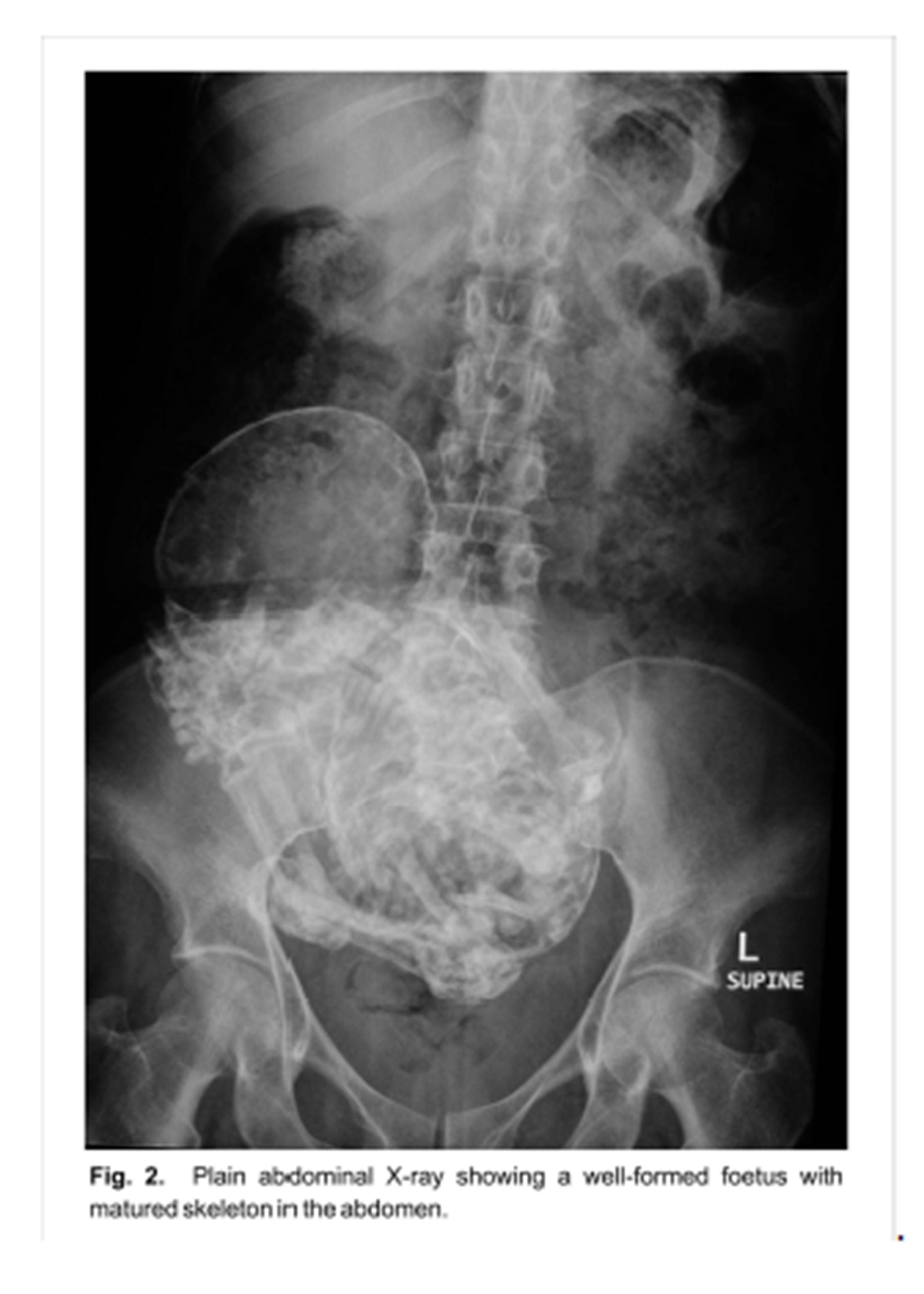
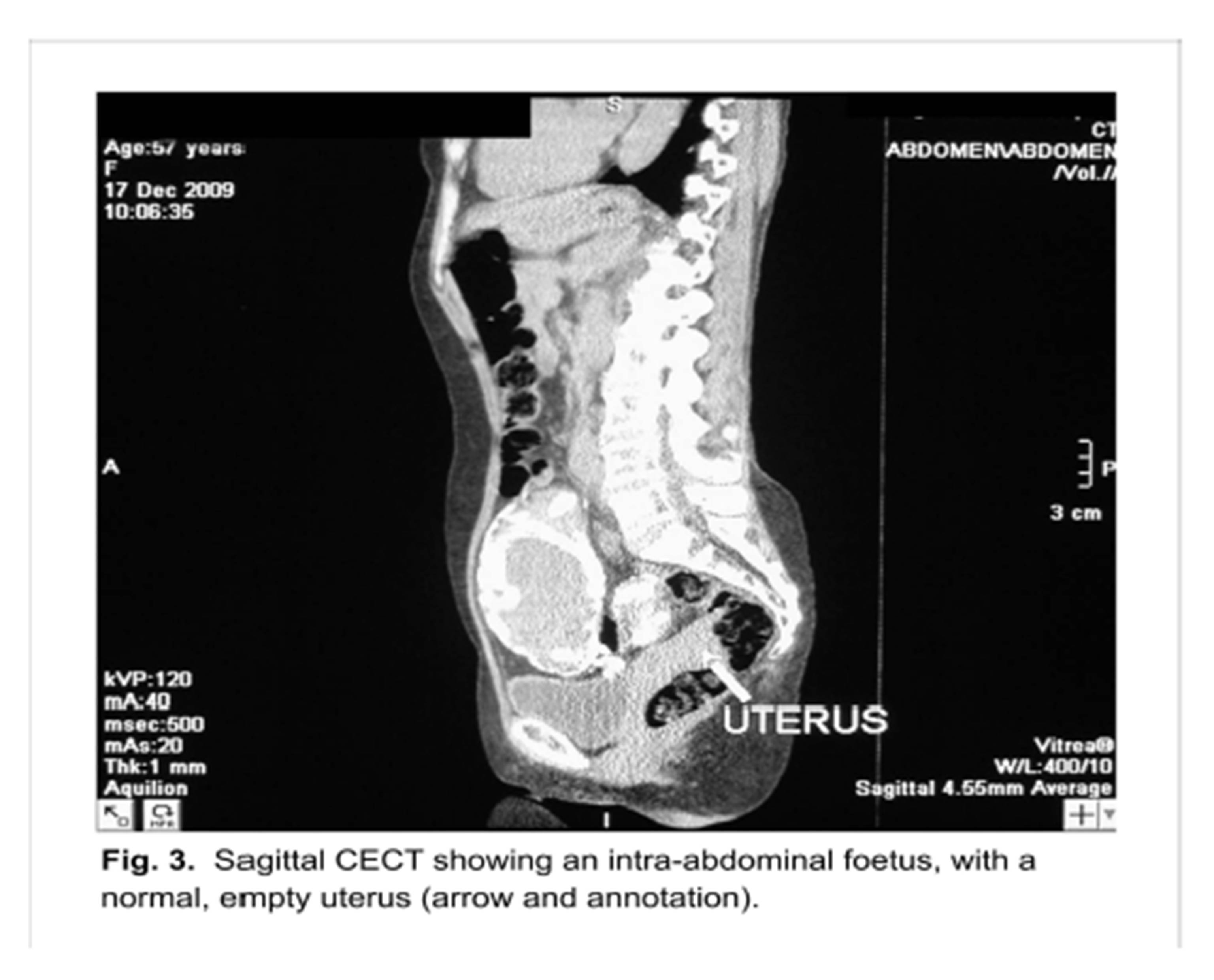
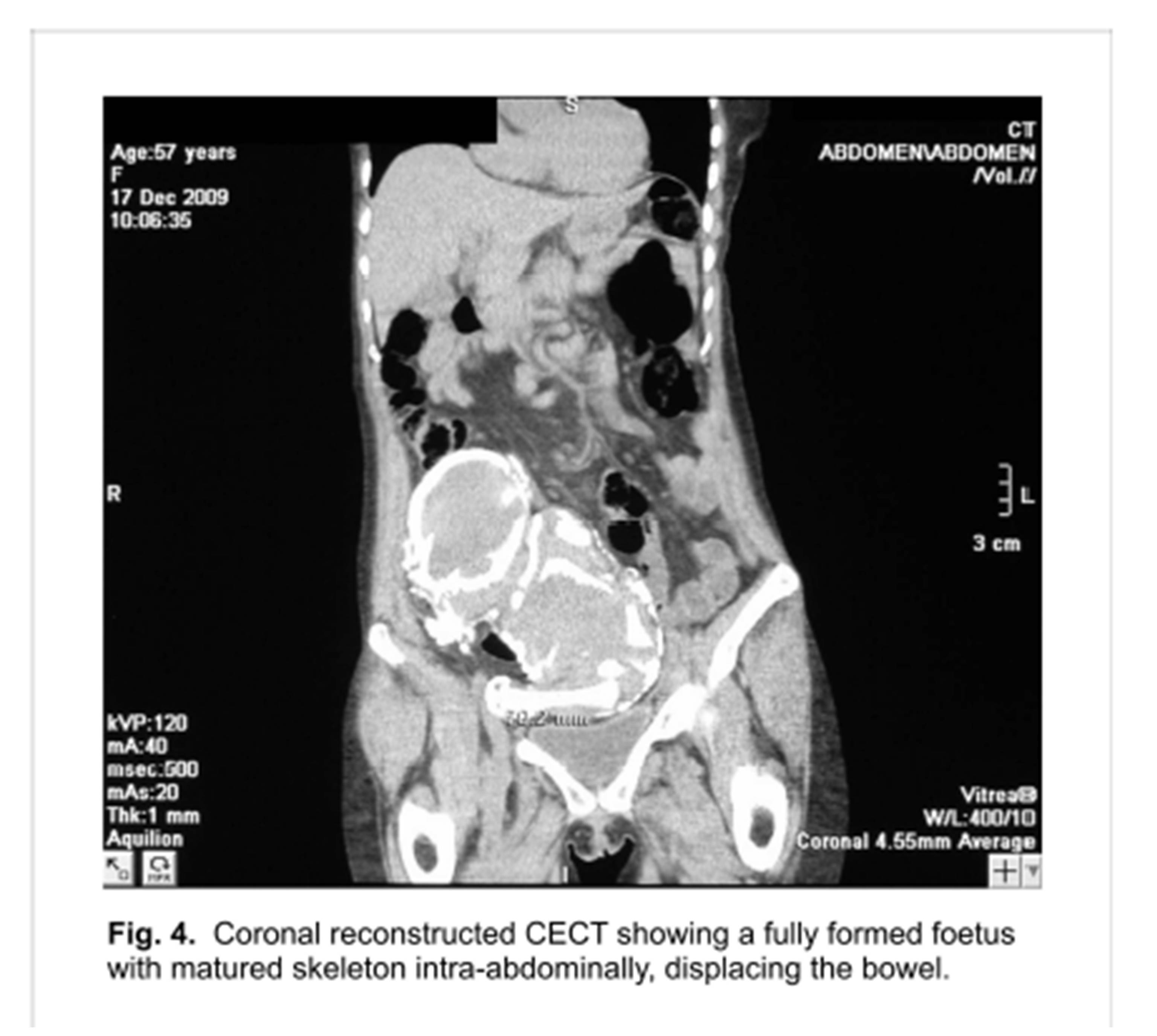
An x-ray of the abdomen is enough to confirm the diagnosis. Computerised tomography (CT scan), and magnetic resonance imaging (MRI) clearly demonstrate the pathology, and assist with the diagnosis of associated complications prior to surgery¹. The CT scan clearly shows the empty uterus and adnexae free of ovular membranes, as in our case.

The differential diagnosis to be considered, especially if the foetus is not clearly defined are teratomas, ovarian tumours, calcified uterine fibroids, inflammatory masses, urinary tract and bladder tumours¹.  In a foetus with well formed skeleton, radiological investigations will reveal the diagnosis of a lithopaedion¹.

Some cases of retained abdominal pregnancy may remain stable without surgical intervention, while others may need early surgical intervention after thorough consideration of the morbidity and the risk of complications if not treated accordingly⁴.

Conclusion

Intra-abdominal pregnancy may be fatal, with significant maternal and perinatal mortality5.
A finding of lithopedion implies an absence of adequate medical attention or some rather serious mistakes in medical judgement2,6.

The case can never over-emphasize the importance of good history taking, systematic and thorough patient examination at all levels of healthcare.  A clinical diagnosis is virtually impossible but radiological investigations like X-Rays, CT-Scan and MRI will clinch the diagnosis.  Discovery of such a condition calls for holistic patient management, considering the psycho-social impact on the patient and family.

In our patient we believe the lithopaedion was not diagnosed earlier because it was not symptomatic and she never sought medical attention for it.  It was an incidental finding in a patient who presented with a hypertensive cerebrovascular accident.

References

1.    Passini JR, Roxana K, Angela PM, et al. Calcified abdominal pregnancy with eighteen years of evolution: Case Report. Sao Paulo Med J/Rev Paul Med 2000;118(6):192-194
 Google Scholar

2.    Lachman N, Satyapal KS, Kalideen JM, et al. Lithopedion: A Case Report. Clin Anat 2001;14:52-54
PublisherGoogle Scholar

3.    Kun KY, Wong PY, Ho MW, et al. Abdominal pregnancy presenting as a missed abortion at 16 weeks’ gestation. HKMJ 2000 Dec;6(4):425-427
Google Scholar

4.    Kim SM, Park S, Lee TS. Old Abdominal Pregnancy Presenting as an Ovarian Neoplasm. J Korean Med Sci 2002;17:274-275
 Google Scholar

5.    Cotter A, Izquierdo L, Heredia F. Abdominal pregnancy [monograph on the internet] The Fetus.net,2000 [2000-10-22-11] Available from: http://www.thefetus.net/page.php?id=1032

6.    Woodbury JW, Jarrett JC. Abdominal lithopedion retained for thirteen years: Case Report with review. Am J Obst & Gynec 1960;80:590-595
 Google Scholar


Shares