Type A Chronic Aortic Dissection in 40-Years Old Smeloff-Cutter Aortic Valve

Antonio Barretta3, Antonino G. M. Marullo1, 2, Mariangela Peruzzi1, Giuseppe Mazzesi3, Piero Proietti3, Ernesto Greco3, David Rose3, Ilaria Chirichilli3, Chiara Santo3 and Giacomo Frati1, 4

1Department of Biotechnology and Medical-Surgical Sciences, “Sapienza” University of Rome, Latina, Italy

2Eleonora Lorillard-Spencer-Cenci Foundation, Rome, Italy

3Department of Cardiac Surgery, “Sapienza” University of Rome, Rome, Italy

4Department of Angiocardioneurology, IRCCS “Neuromed”, Pozzilli (IS), Italy

Academic Editor: Chih-Cheng Lai

Cite this Article as:

Antonio Barretta, Antonino G. M. Marullo, Mariangela Peruzzi, Giuseppe Mazzesi, Piero Proietti, Ernesto Greco, David Rose, Ilaria Chirichilli, Chiara Santo and Giacomo Frati (2014), "Type A Chronic Aortic Dissection in 40-Years Old Smeloff-Cutter Aortic Valve," International Journal of Case Reports in Medicine, Vol. 2014 (2014), Article ID 871865, DOI: 10.5171/2014.871865

Copyright © 2014 Antonio Barretta, Antonino G. M. Marullo, Mariangela Peruzzi, Giuseppe Mazzesi, Piero Proietti, Ernesto Greco, David Rose, Ilaria Chirichilli, Chiara Santo and Giacomo Frati. Distributed under Creative Commons CC-BY 3.0

Abstract

We report the case of a 60-year-old man undergone an aortic valve replacement with a Smeloff-Cutter prosthesis 40 years ago. The patient underwent a redo aortic valve and ascending aorta replacement for ascending aorta aneurysm that intraoperatively appeared as a chronic Type A aortic dissection. The Smeloff-Cutter prosthesis looked intact and functionally normal. The ascending portion of the aorta appeared dissected two centimeters above the sino-tubular junction, between the non-coronary and the left coronary valsalva sinuses: we speculate that Smeloff-Cutter prosthesis may contribute, due to its rheology and features, to the determinism of aortic dilatation and subsequently dissection.
Keywords: Aortic dissection, chronic dissection, smeloff-cutter prosthesis.

Introduction

The Smeloff-Cutter (SC) valve (Cutter Laboratories, Berkeley, CA) was introduced in clinical practice in 1964 and successfully used for almost 20 years. Although less famous than the better-known Starr Edwards (SE) valve (Edwards Laboratories, Santa Ana, CA), the SC prosthesis offered some functional and hemodynamic advantages related to its design. The double cage and the opened struts conformation ensured, in fact, superior flow performances and slightly better long-term results compared to the other first generation ball prosthesis1-3. Few reports have already confirmed the durability of this valve for more than 25 years in aortic position. We describe the case of a patient with an intact 40-year old SC aortic valve and diagnosis of ascending aorta aneurysm.

The Main Body of the Paper

The patient was a 60 year-old man with history of rheumatic disease who underwent aortic valve replacement with a 26-mm Smeloff-Cutter (SC) prosthesis (valve index 16. 2 mm/m2) in 1973 for aortic valve stenosis. Unfortunately, we didn’t find additional pre-operative data concerning the features of the native valve (i.e. tricuspid vs bicuspid). The patient presented to our attention because of recent onset of chest pain, fatigue and shortness of breath. A transthoracic echocardiogram showed normally functioning SC valve with a peak and mean pressure gradient of 19 and 9 mmHg, respectively, and a dilatation of the aortic root and the ascending aorta. The CT scan confirmed the ascending aorta dilatation with a diameter of 58 mm at the level of the aortic bulb, and 71mm at the level of the sino-tubular junction, with images suspicious for aortic dissection (Fig. 1A, B).

871865-fig-1a,1b
The angiography confirmed the aortic dilatation and disclosed a significant stenosis of the left anterior descending artery not providing in any case further information concerning the dissection. Nevertheless, considering these findings, the patient was proposed for surgical treatment. Repair was attempted through a redo median sternotomy after cannulation of the right axillary artery. Once established cardiopulmonary bypass, the ascending aorta was cross-clamped and opened with evidence of an aortic dissection with a completely thrombosed false lumen and an intimal entry site two centimeters above the sino-tubular junction, between the non-coronary and the left coronary valsalva sinuses (Fig. 2).

871865-fig-2

Despite the evidence of a well-preserved and functionally normal prosthesis, considering the anatomical findings and the age of the implant, we decided to replace both, valve and ascending aorta using a n° 27 St. Jude valved conduit. The left anterior descending coronary artery was bypassed with the left internal thoracic artery. The patient was subsequently successfully weaned off from extracorporeal circulation. The explanted prosthesis was evaluated on the bench with no evidence of poppet surface’s abnormalities, ball lipid absorption or pannus deposition around the struts (Fig.3). A specimen of the aortic wall (Fig. 4) was sent for histological characterization. Hematoxylin and eosin staining showed similar morphological characteristics of all the three layers of the aortic walls in both primitive and false lumen (Fig. 4). Negative staining for CD34 and Ki67 markers indicated the absence of cell proliferation, suggestive of a stable structure of the aortic wall, as previously described by our group4, while double staining of endothelium (Pecam-1), and VSMCs (α-SMA) indicated a completed endothelium regeneration in the false lumen confirming that the dissection had overcome its acute phase.

871865-fig-3,4

Discussion

The SC is a first generation heart valve prosthesis that has been successfully implanted in a large number of patients. Functional characteristics of this prosthesis are similar to the Starr Edwards (SE) valve with several important differences related to the structural conformation that determine its unique flow performance making this valve the first “full flow” ball prosthesis. The SC valve has, in fact, an additional titanium cage on the bottom that hold the silicon ball in the valve ring during diastole determining a slight regurgitation around the ball with a consequent self-washing effect that prevents the formation of thrombotic material2. Moreover, the two cages are designed with opened struts limiting the problem of the ball variance, and consenting to use a poppet with a diameter similar to the native valve orifice. The ball is similar to the one adopted in the SE valve and is manufactured using silicon rubber. Even if the lipid absorption by the ball is a possible phenomena, the different silicon’s curing method and the open double caged design of the SC valve partially solved the complications related to the potential ball deformation yielding lower thromboembolism rate and better long-term results1, 2. Different factors have been recognized to positively affect ball-valve durability and freedom from thromboembolic events. Lund et al., identified a valve size index of 13 mm/m2 or less as an incremental risk factor for late mortality, bleeding, and other valve-related complications in patients with SE valve1. Additional factors that have been associated to a longer durability are represented by an age over 50 years at the time of operation, aortic stenosis and ascending aorta dilatation for the favorable effect on pressure gradients and turbulent stress5. Few studies have already confirmed the durability of this valve for more than 25 years in aortic position, and a recent report described an echocardiographic functionally normal SC aortic prosthesis after 40 years6. Despite the evidence of early pannus formation around the struts, thromboembolic events associated with this valve in aortic position have been reported with very low rates, even without permanent anticoagulation therapy,2, 7.

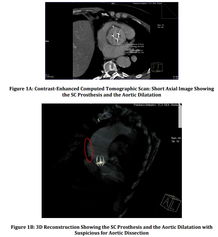
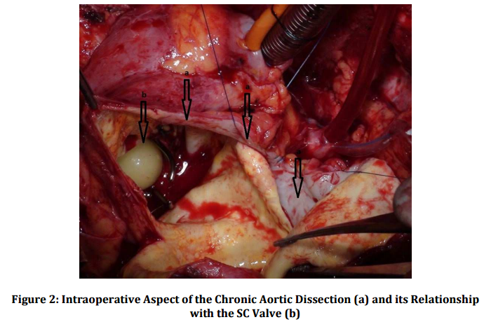
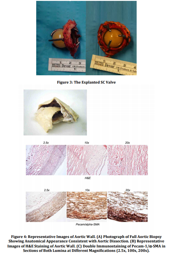
In our case the valve didn’t show structural abnormalities such as ball variance and the poppet appeared smooth with neither alteration of the circular shape nor macroscopic evident lipid absorption. Surprisingly, even if pannus formation around the struts has been reported as a routine complication7, we did not detect either pannus or thrombotic apposition around the valve struts possibly because of the self-washing effect, and the distance of the valve struts from the aortic walls. Blood flow dynamics in ball caged valves might have a significant influence on blood rheology and a great impact in aortic wall degeneration. Thus, it is possible to speculate that flow dynamics imposed by the configuration of the prosthesis causes lateralization of forward flow resulting in blood turbulence and cavitation thus increasing the risk of hemolysis and thromboembolic complications. In our patient moreover, we hypothesized that these lateralized flow characteristics may have contributed to a prolonged increased wall stress, playing a role in the determinism of a progressive aortic dilatation.8  To our knowledge, this case represents the longest reported follow up of functionally normal explanted SC aortic valve, confirming the good features of its ultra-structural conformation, but highlighting that its hemodynamic characteristics were possibly cause of the aortic pathology.

References

1. Lund, O., Pilegaard, H. K., Ilkjaer, L. B., Nielsen, S. L., Arildsen, H. & Albrechtsen, O. K. (1999). “Performance profile of the Starr-Edwards Aortic Cloth Covered Valve, Track Valve, and Silastic Ball Valve,” European Journal of Cardiothoracic Surgery, 16(4):403-13
PublisherGoogle Scholar

2. Stormer, B., Mendling, W., Kohler, J. et al. (1976). “Comparative Study of in Vitro Flow Characteristics between a Human Aortic Valve and a Designed Aortic Valve and Six Corresponding Types of Prosthetic Heart Valves,” EuropeanSurgical Research, 8(2):117-31
PublisherGoogle Scholar

3. Head, S. J., Ko, J., Singh, R., Roberts, W. C. & Mack, M. J. (2011). “43.4-Year Durability of a Smeloff-Cutter Ball Caged Mitral Valve,” The Annals of Thoracic Surgery, 91(2):606-8
PublisherGoogle Scholar

4. Carnevale, D., Lembo, G. & Frati, G. (2011). “Chronic Type A Aortic Dissection: Could Surgical Intervention Be Guided by Molecular Markers?,” Journal of Cellular and Molecular Medicine, 15(7):1615-9
PublisherGoogle Scholar

5. Nygaard, H., Paulsen, P. K., Hasenkam, J. M. et al. (1994). “Turbulent Stresses Downstream of Three Mechanical Aortic Valve Prostheses in Human Beings,” The Journal of Thoracic and Cardiovascular Surgery, 107(2):438-46
PublisherGoogle Scholar

6. Suezawa, T., Morimoto, T., Jinno, T. et al. (2008). “Forty-Year Survival with Smeloff-Cutter and Starr-Edwards Prostheses,” The Annals of Thoracic Surgery, 85(3):14-6
PublisherGoogle Scholar

7. Gometza, B. & Duran, C. M. G. (1995). “Ball Valve (Smeloff-Cutter) Aortic Valve Replacement without Anticoagulation,” The Annals of Thoracic Surgery, 60(5):1312-16
PublisherGoogle Scholar

8. Cohn, L. H. (2007). Cardiac Surgery in the Adult, McGraw-Hill (ed), ISBN: 0071469133
Publisher

Shares