A Post-Vaccination Autoinflammatory Syndrome

Å tefan Rosipal1, Monika Grešíková2, Lukáš Plank3 and Rastislav Rosipal1

1Pediatric Clinic of Preventive Medicine in Poprad, Slovak Health University, Poprad-Veľká, Jarmočná, Slovak Republic/CE

2Clinic of Pediatric Hematology and Oncology, School of Medicine in Bratislava, Comenius University, Slovak Republic/CE

3Department of Pathology, Jessenius School of Medicine in Martin, Comenius University, Slovak Republic/CE

Academic Editor: Katsuiku Hirokawa

Cite this Article as:

Å tefan Rosipal, Monika Grešíková, Lukáš Plank and Rastislav Rosipal (2014), "A Post-Vaccination Autoinflammatory Syndrome," Research in Immunology: An International Journal, Vol. 2014 (2014), Article ID 537063, DOI: 10.5171/2014.537063

Copyright © 2014 Å tefan Rosipal, Monika Grešíková, Lukáš Plank and Rastislav Rosipal. Distributed under Creative Commons CC-BY 3.0

Abstract

The current massive vaccination brings a Janus- faced problem. There is a need to assess a positive effect but also to anticipate the abnormal immune response. Repeated administration of aluminum containing vaccines represents a possible risk factor for different aberrant reactions including the autoinflammatory syndrome that at this stage can be called enigmatic post-vaccination phenomena. Described case gave rise to the suggestion that vaccination could cause the undesired and unintentional hyper activation of the inflammatory system due to lymph node hyperplasia. Castleman like disease is presented as autoinflammatory syndrome. The dysfunction in the immune/inflammatory response seems to play an etiologic role and leads to a high degree of systemic, sterile inflammation. A temporal relationship between vaccination and the adverse reactions, latency period, can involve weeks and months. Autoinflammatory disease is interesting by severe chronic anemia of inflammation and with decreased levels in serum lipid profile. The extirpation of localized lymphoid tumor in the abdomen was beneficial to the patient and immediately ended-up the inflammatory state and normalized the abnormal laboratory results. Studies addressing this issue are limited. 

Keywords: Vaccination, autoinflammatory syndrome, lymph node hyperplasia, Castleman disease

Introduction

Vaccination has been the most successful medical intervention in history. At present it is in children and adolescents very effective tool to prevent infectious diseases. On the other hand, vaccination evokes a problem of increasing fear of post-vaccine adverse reactions in previously healthy individuals, Siegrist (2007). An activated immune/inflammatory system is tightly regulated process and the pathogenesis of undesirable events has been yet not fully elucidated. Auto inflammatory disease, syndrome is a classification for chronic inflammation with a distinct therapeutic role for blocking pro-inflammatory cytokines, especially interleukin-1β. A coherence of vaccination and autoimmune/inflammatory syndrome is formulated mainly on the basis of case reports and the causal link is vehemently disputed. These diseases arise in genetically predisposed subjects and require an environmental trigger including vaccination, Wraith et al (2003). The latency period can take time weeks or month-long, Agmon-Levin et al (2009).The type associated with vaccination is entitled as ASIA- Autoimmune/inflammatory Syndrome Induced by Adjuvants, in a study by Shoenfeld et al (2011). Diagnosing a new patient that supports the causality between vaccination and presentation of aberrant response needs a precise history and differential diagnosis.  

Case Report

Medical history and physical exam: a 21-year-old man with normal growth and development.  He has been subject to the routine vaccination according to the Slovak vaccine schedule. Family history was uneventful. Patient suffered from a mild signs of pollen allergy at preschool time. At age 13 hepatitis A, B vaccine and tetanus toxoid (all contain aluminum) were administered in the recommended intervals. One year ago adolescent had physiological complete blood count including hemoglobin (139-141g/L), erythrocyte count (4.44-4.87 10*12/L), hematocrit (45-46%) and erythrocyte sedimentation rate (9/21, 10/21). Physical examination, blood pressure, pulse, weight and height were unremarkable. At age 17 by a regular preventive medical care mild hypochromic anemia was detected along with high grade of systemic inflammation. Patient had no clinical signs. That time began the large clinical and laboratory studies. Subhepatic tumor was depicted by ultrasound and computer tomography (CT), size 60x 44x 40 mm. A probative laparatomy was performed in order to obtain the samples of affected subhepatic and paracaval lymph node- incisional biopsy. The histological finding was incomplete for diagnosis of Castleman disease, no malignant cells. The conclusive diagnosis remains problematic. A series of intravenous iron medicament were administered but without an expected effect. At the age of 18 the patient visited our outpatient department. Only occasional fatigue and dry mouth appeared in clinical manifestation. The differential diagnosis was focused on a broad spectrum of the pediatric inflammatory diseases that were analysed prior to the acceptance of the adverse reaction linked to vaccination. We suggested an postvaccinal autoinflammatory syndrome. Because of the administrative problem there was no opportunity of applying the human monoclonal antibody, inhibitor of interleukin- 1β (canakinumab). At age 19 a pharmacotherapy was started with Prednisone at a dose 60mg per day, but the treatment was complicated with recurrent thrush. Glucocorticoid didn’t modify the abnormal laboratory results. Despite the previous non-malign biopsy, the second surgical intervention was indicated with the aim of a radical cure. Four transfusion unit of red blood cells and two products of fresh frozen plasma were transfused before the laparatomy. The retroperitoneal tumor was completely resected and perhaps a combination with biological preparations administered intravenously caused a rapid and sustained cessation of symptoms, and also the normalization of the abnormal biochemical and hematological markers.     

The Abnormal Results (Reference Ranges)

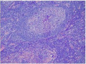
Hemoglobin 72-110g/L (140-180), hematocrit 0.293-0.394 (0.420-0.520), MCV 62.1- 79.0 fl (82-100), MCH 18.9-23.8 pg (27.0-33.0), MCHC 285-338 g/L (330-370), ESR 66-120/ 90-160 (9/21); CRP 75.2-156.2 mg/L (0.0-5.0), procalcitonin 0.32-1.11 ng/L (<0.5), iron 1.98-2.7 µmol/L (10.6-28.3), inadequate iron absorption, copper 32.12-38.00 µmol/L (11.0-24.0), erythropoietin 28.3-42.3 IU/L (3.5-17.6), haptoglobin 5.55 g/L (0.50-3.20), fibrinogen  6.20-11.29 g/L (2.00-4.50), transferrin 1.80-2.33 g/L (2.00-3.60), soluble transferrin receptor 2.31-11.03mg/L (1.30-5.0), serum hepcidin (ELISA) 63.9 µg/L relatively elevated level (29-254). Trephine bone marrow biopsy: mild hypercellularity of the hematopoietic bone marrow (BM) with proportionally distributed precursors of all BM cell lineages. The maturation is trilineary preserved with focally accented macrocytic dyserythropoiesis, no dysplastic changes and stroma fibrosis. The iron reserves are reduced. Alpha-1-antitrypsin 2.27-3.10 g/L ((0.90-2.00), IgG 27.58-31.20 g/L (7.00-16.00), IgG1 13.789-15.395 g/L (4.900-11.400), IgA 4.20-4.34 g/L (0.70-4.00), IgA1 5012.00mg/L (613.00-3040.00),  IgE 779-811 IU/ ml (<100), IgD 391mg/L (<140.0) total protein 83.5-87.5 g/L (66.0-83.0), albumin 31.8-35.6g/L (32.0-52.0), electrophoresis fractions: albumin 40.80% (58.0-65.0), alpha 2 globulin 14.7% (5.0-10.0) beta 2 globulin 5.40% (2.50-4.50), gamma globulin 28.30% (10.0-19.0), OGTT- fasting glucose 6.10mmol/L (<5.60mmol/L), HbA1c 5.80%/39.9mmol/mol (<5.50%/<37.00mmol/mol), trend to insulin resistance, total cholesterol 2.56-3.15mmol/L (3.30-<4.25), HDL cholesterol 0.56-1.11mmol/L (1.10-<1.55), LDL cholesterol 1.51-2.15mmol/L (1.60-<2.65), triacylglycerols 0.41-0.60mmol/L (0.53-<1.15), apolipoprotein B 0.54-0.61g/L (0.60-<0.85), apolipoprotein A-I 1.10-1.13 g/L (1.15-<1.70), interleukin-6 (IL-6) 10.89-51.55 pg/ml (0.00-7.00). Ultrasound, CT, magnetic resonance imaging (MRI) of the abdomen have showed retroperitoneal isolated tumor. Its description by positron emission tomography (PET) combined with CT: increased metabolism of glucose in the viable lesion. This solitary hypodense, non penetrans tumor was located in the subhepatic space under the left lobe of hepar, size 61x46x39mm. Operative procedure confirmed the solid tumor retroperitoneal retrohepatalis, next to the arteria hepatica propria and vena gastrica sinistra, size 80x60x50mm. Exstirpatio tumoris, and lymphadenectomia tractus coeliacus. Excisional biopsy: the micrographs show features of chronic antigenic stimulation of the lymphatic parenchyma with predominance of advanced reactive follicular hyperplasia, perifollicular and medullary plasmacytosis (Fig.1). The plasma cells are immunohistochemically polyclonal and some of the hyperplastic follicles present transition to the regressive phase of the hyperplasia with increased germinal center cells apoptosis and expansion of the marginal zone. In contrast to bcl2 negative germinal centers, the lymphoid cells of the follicular and interfollicular compartment showed immunohistochemical positivity of bcl2 protein. There are small not extensive foci of hyalinosis in the stroma, no sign of amyloidosis. The criteria for the biopsy diagnosis of Castleman disease, plasma cell type of angiofollicular hyperplasia, were not fulfilled. The amount of aluminum in the tumor tissue was 7µg/g, seven fold more than the control.

537063-fig-1

Figure 1. Chronic Antigenic Stimulation of the Lymphatic Parenchyma. The Reactive Follicular Hyperplasia, Advanced Perifollicular and Medullary Accumulation of the Mature Plasma Cells (Plasmacytosis)

 
The Normal Results

RBC 5.00-5.40 10*12/L (4.30-5.70), leukocytes and their distribution, platelet count, reticulocytes, ferritin, interleukin- 1β (IL1-F2) 0.65 pg/ml (0.001-2.5), IgM, IgG2, IgG3, IgG4, IgA2, minerals, ceruloplasmin, urin analysis, complement C3 and C4, paraproteins, ASLO, uric acid, repeated glucose, fasting serum insulin level 42.38pmol/L (<56.00), vitamin B12, folic acid, lipoprotein (a) 0.14g/L (<0.20), thomocystein 10.3µmol/L (4.0-15.4). Renal, liver, pancreatic and gastroenterological (including malabsorption) sets, infection verification (causes), hematologic tests without anemia, autoimmune diseases and oncological set, no endocrinopathies. Imaging tests: the organs and cavities, neck by ultrasound, CT, MRI, PET/CT not detected another enlarged lymph nodes. In plasma: IL-1α, IL-2, IL-4, IL-10, IL-12/23, IL-17A, TNF alpha, IFN gamma, Th1/Th2 ratio. Mutational analysis: JAK2V617F, factor V Leiden (G1169A), factor II prothrombin (G20210A), MTHFR (C677T, A1298C)

Postoperative Results

The recheck in three weeks: ESR 10/34, RBC 5.47 x10*12/L, hemoglobin 127 g/L, hematocrit 0.388, MCV 71.00 fl, MCH 23.2 pg, MCHC 327.0 g/L, reticulocytes 0.72%, iron 10.2 µmol/L, transferrin 3.23 g/L, soluble transferrin receptor 6.65 mg/L, ferritin 141.59 µg/L, flow cytometry, fibrinogen 2.8 g/L, CRP 2.8 mg/L, interleukin-6 3.2pg/ml, procalcitonin 0.06 ng/ml, total cholesterol 5.00 mmol/L, HDL cholesterol 1.18 mmol/L. The recheck in twelve weeks: ESR 6/20, RBC 5.50×10*12/L, hemoglobin 13.6g/L, hematocrit 0,435, reticulocytes 0.93%, iron 16.2µmol/L, s. transferrin receptor 2.9 mg/L, fibrinogen 3.1 g/L, CRP 2.5mg/L, total cholesterol 3.21 mmol/L, HDL cholesterol 1.09 mmol/L, LDL cholesterol 1.57 mmol/L, triacylglycerols 0.55 mmol/L, interleukin-6  2.1 pg/ml,  IgG 13.55g/L, IgG1 7.85g/L, IgA1 2120mg/L,  IgD 90mg/L, IgE 115 IU/ml.    

Discussion

The inflammation is a complex biological reaction to pathogenic and harmful stimuli. Autoinflammatory syndrome currently represents about nineteen different diseases. They are primarily and mainly mediated by interleukin- 1 family, especially IL-1β, Dinarello (2009). Clinical features, laboratory studies, genetics excluded the pediatric hereditary inflammatory syndromes, and other well-known immunological and inflammatory dysfunctions too. The exposure to an external stimuli (vaccine, adjuvant) prior to clinical manifestation, chronic fatigue and dry mouth were observed in patient according to the suggested major criteria for the diagnosis of ASIA. The new diseases including macrophagic myofasciitis, postvaccination phenomena and Gulf War Syndrome may be incorporated into autoimmune/inflammatory syndrome induced by adjuvants (Shoenfeld`s syndrome). A fever is not a striking feature of the autoinflammatory syndrome. Castleman disease must be taken into consideration by the differential diagnosis of well- defined enhancing mass in the abdomen, its unicentric type. Retroperitoneal localization is exceptional. The etiology is not known for sure. Diagnostic clue to this disease is an elevated concentration of IL-6 in plasma, chronic, iron-refractory mikrocytic anemia and positive PET/CT scan. The diagnosis should be confirmed by histological examination, and traditionally three histological variants are distinguished.  Castleman disease is closely mimicked by IgG4-related sclerosing disease, in Jo et al (2011). The conclusive diagnosis in our patient was Castleman like disease that is presented as autoinflammatory syndrome. Vaccination can be other possible cause of lymph node hyperplasia, adjuvant related disease. It is very difficult exactly to determine the beginning of post-vaccination progressive process of the inflammation, because the clinical long-lasting latency period delays the indication of the laboratory studies and imaging tests. The process might start immediately post vaccination. Aluminum adjuvant stimulates Th2 type response and increases the synthesis of cytokines (IL-1, IL-4) as well as B/plasma cells production of IgG1, IgA1, IgD and IgE, in research study by Brewer et al. (1997). Aluminum alone has several pathologic effects such as impact on the erythropoiesis and is the agonist of the inflammasome- caspase cascade. The deposition of aluminum into tumor formation is one of the pathomechanisms, which occurs in this clinical observation. The determination of the non-physiological level of aluminum in patient`s lymphatic tumor is still unresolved and data about its amounts are difficult to interpret. The information retrieval may shed new light on the potential immunotoxicological role of aluminum- adjuvanted vaccines, Salemi et al (2010). Interleukin-1β along with interleukin-6 stimulate the induction of acute- phase response with subsequent production of the corresponded reactants by hepatocytes. The elevated levels of the interleukin-6 promote the large spectrum of clinical and mainly laboratory abnormal findings. Its increased concentrations cause an amplification of the inflammatory process and also accelerate the maturation of the plasma cells. Fibrinogen is a marker of inflammation rather than hemostasis. During the acute phase response serum levels of high-density lipoproteins drop via increased enzymatic hydrolysis of the lipoprotein. It is chiefly the effect of the endothelial lipase and secreted phospholipases A2. The inflammation causes a chronic anemia at different levels of erythropoiesis. The overproduction of hepcidin or its relatively high activity according to anemia impinges iron traffic leading to retention of the metal in the competent cells and thus to iron deficient erythropoiesis, Bergamaschi et al (2009).  Interleukin-6 is the major inflammation- driver inducer of hepcidin synthesis and is also involved in differentiation of hematopoietic precursor cells, Nemeth et al (2004). The linkage between hepcidin and ferroportin blocks the transfer of iron from the duodenal mucosal cells into the circulation. This link increases the retention of iron within macrophages-inhibited recycling and deteriorates reutilization of iron by erythrophagocytosis. The pro-inflammatory cytokines have inhibitory effects on erythroid progenitor cells and reduce the expression of hematopoiesis growth factors, Weiss et al (2010). Alpha-1 antitrypsin inhibits the iron uptake into the erythroid progenitors.  

Taken together, vaccines administration with aluminum adjuvant might be a potent stimulating factor on the immune/inflammation system that sometimes launches non-infectious systemic inflammation. Aluminum containing vaccines could initiate and promote the lymphoproliferative disease. This process enhances the synthesis and release of bioactive substances that provoked autoinflammatory syndrome. Blunting of the biological activity of erythropoietin along with the failure to properly utilize iron are the major causes of chronic anemia of inflammation. The pathogenesis of the post-vaccination phenomena is not completely understood, as well as its long-term health consequences.  The vague symptoms and physical findings cause that in clinical practice the autoinflammatory syndrome is often not in time categorized into the accurately defined disease. The extirpation of the lymphoid tumor activating pro-inflammatory condition is the adequate kind of the treatment. In this patient follow-up exams are recommended for several years after the treatment was finished. More research and much greater knowledge are needed to clarify the relationship between aluminum adjuvant and the development of the autoinflammatory syndrome. 

References

Agmon-Levin, N., Paz, Z., Israeli, E. & Shoenfeld, Y. (2009). “Vaccines and Autoimmunity,” Nature Reviews Rheumatology, 5 (11) 648-652.
PublisherGoogle Scholar

Bergamaschi, G. & Villani, L. (2009). “Serum Hepcidin: A Novel Diagnostic Tool in Disorders of Iron Metabolism,”Hematologica, 94 (12) 1631-1633.
PublisherGoogle Scholar

Brewer, J. M, & Alexander, J. (1997). “Cytokines and the Mechanisms of Action of Vaccine Adjuvants,” Cytokines, Cellular & Molecular Therapy, 3 233-46.
PublisherGoogle Scholar

Dinarello, C. A. (2009) “Immunological and Inflammatory Functions of the Interleukin-1 Family,” Annual Review of Immunology, 27 519-50.
PublisherGoogle Scholar

Jo, J.- H., Park, Y.- S., Jeon, Y. K., Nam, S. J. & Huh, J. (2011). ”Comparison of Plasma Cell Type of Castlemans Disease and IgG4-Related Sclerosing Disease: A Histopathological and Immunohistochemical Study,” Pathobiology 78(4) 227-32.
PublisherGoogle Scholar

Molina, V. & Shoenfeld, Y. (2005). “Infection, Vaccines and Other Environmental Triggers of Autoimmunity,”Autoimmunity 38 (3) 235-245.
PublisherGoogle Scholar

Nemeth, E., Tuttle, M. S., Powelson, J., Vaughn, M. B., Donovan, A., Ward, D. M., Ganz, T. & Kaplan, J. (2004). “Hepcidin Regulates Cellular Iron Efflux by Binding to Ferroportin and Inducing its Internalization,” Science, 306 (5704) 2090-2093.
PublisherGoogle Scholar

Salemi, S. & D’Amelio, R. (2010). “Vaccination and Autoimmune Diseases,” International Reviews of Immunology, 29 (3) 239-246.
PublisherGoogle Scholar

Shoenfeld, Y. & Agmon- Levin, N. (2011). “‘ASIA’- Autoimmune/Inflammatory Syndrome Induced by Adjuvants,” Journal of Autoimmunity, 36 (1) 4-8.
PublisherGoogle Scholar

Siegrist, C.- A. (2007). “Autoimmune Diseases after Adolescent or Adult Immunization: What Should Expect?,” Canadian Medical Association Journal,  177 (11) 1352-1354.
PublisherGoogle Scholar

Weiss, G. & Gasche, C. (2010). “Pathogenesis and Treatment of Anemia in Inflammatory Bowel Disease,” Hematologica, 95 (2) 175-178.
PublisherGoogle Scholar

Wraith, D. C., Goldman, M. & Lambert, P.- H. (2003). “Vaccination and Autoimmune Disease: What is the Evidence?,”Lancet, 362 (9396) 1659-1666.
PublisherGoogle Scholar

Shares