Isolation of a Sequence Homolog to More Axillary Branches MAX2 Gene in Hibiscus rosa-sinensis and its Use as Genetic Marker

Luca Braglia1, Federica Nicoletti2, Laura De Benedetti3, Nicola Pecchioni4 and Antonio Mercuri3

1CNR-IBBA Institute of Agricultural Biology and Biotechnology, Milano, Italy

2Consiglio per la Ricerca e la Sperimentazione in Agricoltura – Centro di Ricerca per le Colture Industriali (CRA-CIN), Bologna, Italy

3Consiglio per la Ricerca e la Sperimentazione in Agricoltura – Unità di Ricerca per la Floricoltura e le Specie Ornamentali (CRA-FSO), Corso Inglesi, Sanremo (Imperia), Italy

4University of Modena and Reggio Emilia, Department of Agricultural and Food Sciences, Reggio Emilia, Italy

Academic Editor: Jan De Riek

Cite this Article as:

Luca Braglia, Federica Nicoletti, Laura De Benedetti, Nicola Pecchioni and Antonio Mercuri (2015), "Isolation of a Sequence Homolog to More Axillary Branches MAX2 Gene in Hibiscus rosa-sinensis and its Use as Genetic Marker," Research in Agriculture and Agronomy, Vol. 2015 (2015), Article ID 360263, DOI: 10.5171/2015.360263

Copyright © 2015. Luca Braglia, Federica Nicoletti, Laura De Benedetti, Nicola Pecchioni and Antonio Mercuri. Distributed under Creative Commons CC-BY 4.0

Abstract

Lateral branching plays an important role in the elaboration of adult plants architecture. Herein, we adopted a modified AFLP approach combined with a degenerate primer amplification to identify and isolate in the underinvestigated ornamental species H. rosa-sinensis an orthologous element of the MAX2 gene (More Axillary Branches), which acts downstream of the branching inhibition signaling pathway. A specific gene fragment was cloned and sequenced from nineteen H. rosa-sinensis cultivars and twelve Hibiscus botanical species and different significant nucleotide polymorphisms among genotypes that were observed. The comparative analysis revealed a high conservation of DNA sequences among cultivars and wild species sexually compatible with H. rosa-sinensis. The deduced amino acid sequences of the Hibiscus isolated fragments reveal four characteristic repeat regions showing high identity with other F-box/Leucine Reach Repeat MAX2 homologous sequences. The cloned fragment is a likely candidate gene to be validated for association with phenotype to release a gene-derived “perfect marker” for the compact habit trait.

Keywords: Hibiscus – AFLP approach – ornamentals – shoot branching.

Introduction

Hibiscus rosa-sinensis is one of the most widely planted ornamental shrubs cultivated throughout the tropics and sub-tropics. Numerous varieties and hybrids are particularly appreciated in garden and landscape for their vigorous growth habit, but they are mainly employed as a pot plant when growth retardants for keeping a reduced plant size are applied. A compact basal branching growth habit is generally preferred.

Lateral branching structures exist in many forms throughout higher plants and even if plant architectures are influenced by environmental factors, their species-specific characteristics indicate the presence of widely conserved genetic regulatory mechanisms (McSteen and Leyser 2005, Johnson et al 2006).

In different plant species, pre-existing axillary meristems may either lie dormant for long periods or they may develop into branches instantaneously. This bud growth can be activated by intrinsic factors, and hormones play a crucial role in shoot branching control (Leyser 2009). It has been known for many years that auxin synthesize in the apex inhibits axillary meristems outgrowth, whereas cytokinin promote it efficiently by regulating the shoot branching phenomena (Liang et al 2010).

However, the involvement of a novel hormone-signaling pathway in the regulation of bud growth has been inferred by genetic analyses of mutants that have enhanced shoot branching phenotype in Arabidopsis, pea, petunia and rice (Booker et al 2005, Morris et al 2001, Snowden and Napoli 2003, Arite et al 2009). In Arabidopsis thaliana, a suite of mutants with More Axillary Branches encoded by the MAX1, 2, 3, and 4 genes has been analyzed by a combination of grafting and molecular techniques (Bennet et al 2006). The recessive mutations (max) cause premature and enhanced outgrowth of lateral shoots in combination with modest pleiotropic effects. These studies suggested that MAX1, 3 and 4 are involved in the synthesis of a mobile signal, whereas the MAX2 gene product mediates perception and response to the signal (Leyser 2009). Particularly, MAX2 has been shown to encode a nuclear localized F-box leucine-rich repeat (LRR) protein within the SCF (Skp1-Cullin-F box) complex that catalyzes the ubiquitination of proteins, and thus target them for proteasomal degradation (Xu et al 2009). In the case of MAX2, one or more proteins that activate bud growth are in the wild-type targeted for destruction by the MAX2 F-box LRR product (Stirnberg et al 2007). Presumably, these proteins, which would be stabilized in the absence of MAX2 activity, would in some way promote branching.

More recently, Wang et al (2013) demonstrated in Arabidopsis that the strigolactone hormone inhibits auxin transport, suggesting a complex interaction between these two hormones and the MAX2 F-box binding site in the protein degradation system.

Given the metabolic complexity of plants, there are probably more, perhaps many more, small molecules with signaling function. However, the discovery of regulatory mechanisms promoting the axillary branch proliferation could provide an environmentally independent, rapid and helpful tool for preliminary screening of genotypes characterized by a compact basal branching growth habit, suitable for pot plant cultivation.

With the aim to identify a gene-derived ‘perfect’ molecular marker associated to the compact plant architecture, we isolated conserved sequences for the MAX2 gene in the underinvestigated ornamental species Hibiscus rosa-sinensis. The knowledge gained through the previous AFLP characterization of a collection of H. rosa-sinensis cultivars (Braglia et al 2010) allowed us to develop a new strategy. Starting from plant MAX2 gene sequences, we followed a combined approach of degenerate primer PCR together with AFLP technique

Material and Methods

Genomic DNA was isolated from nineteen Hibiscus rosa-sinensis cultivars with different and contrasting plant architectures and thirteen Hibiscus botanical species (H. arnottianus G., H. boryanus H. and A,. H. calyphyllus Cav., H. cannabinus L., H. denisonii B., H. genevii B., H. kokio H., H. moscheutos L., H. panduriformis B., H. schizopetalus H.,H. storckii S., H. syriacus L., H. tiliaceus L.), selected from materials collected at the CRA-FSO in Sanremo (Italy). DNA from one hundred milligrams of fresh leaves was extracted using the DNeasy Plant Mini Kit (Qiagen, Germany) following the modified protocol reported for Hibiscus spp. by Braglia et al (2010).

According to the full-length cDNA sequence of Arabidopsis MAX2 gene (NM_129823), pea RAMOSUS4 gene (DQ403159), rice LRR-repeat MAX2 homolog (Oryza sativa Japonica group) (Q5VMP0) and poplar F-box family protein mRNA sequence (Populus trichocarpa) (XM_002320376) showing strong homology at the amino acid level, a consensus sequence by multiple sequence alignment was generated. A set of six degenerate primers were then designed and tested on three different Hibiscus rosa-sinensis genomic DNAs. PCR reactions were performed in a 50 µl containing 1X PCR Buffer (HotStartTaq®Plus Buffer Qiagen, Germany), 0.2 mM each dNTP, 2 mM MgCl2, 150 ng of DNA template, 1.6 µM primer and 2.5U Taq DNA Polymerase (HotStartTaq®Plus Qiagen, Germany). The PCR conditions were 2 min at 94° C, 5 cycles 30 s at 94° C, 45s at 48° C, 2 min at 72° C, followed by 35 additional cycles 30 s at 94° C, 45 s at 58° C, 90 s at 72°. The reactions were held at 4°C after a final extension at 72°C for 10 min.

The amplified products were separated using agarose electrophoresis (2.2 %). Only one primer pair (dF3 5’-TTYACNGARGGNTTCAAGTC-3’; dR2 5’-CCYTGRAAGTGNCCNAGCTT-3’) yielded the expected size fragment. This PCR product was subsequently cloned (TA Cloning® kit, Invitrogen) and sequenced, then analyzed using bioinformatic tools at the websites http://www.ebi.ac.uk/Tools/ and http://www.ncbi.nlm.nih.gov/.

Hibiscus specific primers (notable as Hsp_, Table 1) were designed on the isolated fragment sequence in forward and reverse. An AFLP (Amplified Fragment Length Polymorphism)-based approach was adopted to extend the Hibiscus DNA segment outside the boundary known sequence. Restricted/ligated fragments (EcoRI/MseI), hereafter R/L were generated according to the AFLP protocol reported by Vos et al (1995) from 300 ng of genomic DNA. The obtained R/L products were used to test different Hsp_ primers in combination with AFLP primers (Table 1). These latter, named Ead_pr and Mad_pr, had the 5’-region complementary to the adapter and the restriction site sequence without selective nucleotides at the 3’-end.

PCR reactions were performed in a 25 µl containing 1X PCR Buffer (HotStartTaq®Plus Buffer Qiagen, Germany), 1 µl R/L, 0.2 mM each dNTP, 1.5 mM MgCl2, 0.5 µM for the Hsp_ primer and 0.1 µM for the AFLP primer with 2.5U of Taq DNA Polymerase (HotStartTaq®Plus Qiagen, Germany). The following PCR conditions were used: 5 min at 95 °C, 13 cycles of 30 s at 90°C, 30 s at 67°C, 60 s at 72°C with a decrease of 0.7°C of the annealing temperature carried out in each cycle followed by 27 additional cycles of 30 s at 90° C, 30 s at 56° C, 60 s at 72°. The reactions were held at 4°C after a final extension at 72°C for 10 min. The obtained PCR product was subsequently cloned (TA Cloning® kit, Invitrogen) and sequenced. One hundred additional base pairs were achieved from the AFLP-based approach and two new Hsp_ reverse primers were synthesized (Fig. 1, Table 1).

Hibiscus MAX2 Gene Fragment Schematic Representation. MseI Restriction Site is Reported at the 5’ Region. Black Arrows Indicate the Name and Position of Hsp_ Specific Primers.

 Fig. 1: Hibiscus MAX2 Gene Fragment Schematic Representation. MseI Restriction Site is Reported at the 5’ Region. Black Arrows Indicate the Name and Position of Hsp_ Specific Primers.

Table 1: Hibiscus Specific Primers and AFLP Core Primers

360263-TABLE-1

The selected primer pair Hsp_Up1Hsp_Dw1 was tested on all genomic samples using the same PCR conditions reported above. Amplified fragments were sequenced to assess nucleotide polymorphisms.

Cluster analysis of Hibiscus MAX2-like isolated nucleotide sequences was performed using the Treecon program for Windows (Van de Peer and De Wachter 1994), with a bootstrap test (Hillis and Bull 1993).

The amino acid sequences were deduced and the sequence comparison was conducted through database search using UniProt (Universal Protein Research http://www.uniprot.org)

Results

The degenerate primer amplification allowed the identification of a DNA fragment (~350 bp) showing 65% similarity to the Arabidopsis MAX2 gene and the deduced amino acid sequence revealed a high degree of homology with those of other F-box subunit proteins from various biological sources: Populus trichocarpa (64%), Arabidopsis thaliana (64%), Pisum sativum (61%) and Oryza sativa (32%), most of them employed in the degenerate primer design.

Concerning to the AFLP approach, all tested specific primers in combination with the primer Ead_pr did not produce any amplification products (data not shown). Whereas, the primer combination Mad_pr/Hsp_Up1 allowed to extend theHibiscus MAX2-like sequence downstream the 3’ boundary known sequence of one hundred additional nucleotide base pairs. Unfortunately, the presence of a MseI restriction site at the 5’ region of the isolated fragment (named HibMAX2-like) did not permit to extend the sequence upstream (Fig.1).

The HibMAX2-like sequences could be amplified in all cultivars of Hibiscus rosa-sinensis and Hibiscus species except for H. cannabinus (kenaf). Control reactions on genomic DNA samples of Arabidopsis and pea did not produce any amplicons (Data not shown). The Hibiscus MAX2-like nucleotide sequences were submitted to NCBI database with the Accession Numbers JF813799-JF8137824. A neighbour-joining tree was then constructed (Fig. 2) for all Hibiscus isolated nucleotide sequences. In this tree, a main cluster (A, bootstrap value 98%) could be recognized, grouping all Hibiscus rosa-sinensis cultivars. Furthermore, some botanical species such as H. arnottianus, H. boryanus, H. denisonii, H. genevii, H. kokio, H. schizopetalus and H. storckii were spread throughout the main cluster. Conversely, H. calyphyllus, H. moscheutos, H. panduriformis, H. syriacus and H. tiliaceus, were grouped in separate clusters.

 360263-fig-2

Fig. 2: Neighbor-joining Tree Built from HibMAX2 Nucleotide Sequences of 19 Hibiscus rosa-sinensis Varieties and 12 Hibiscus Botanical Species. The Arabidopsis MAX2 Sequence was Reported as an Outgroup. Numbers on Nodes Indicate the Bootstrap Values after 1000 Replicates.

Although the deduced HibMAX2-like amino acid sequences lack the conserved N-terminal and C-terminal domains, the comparison of these sequences revealed the presence of the characteristic repeat regions in all samples analyzed (black boxes in Fig. 3). Indeed, in silico analysis showed the presence of four LRRs of the motif LxxLxL, with L (leucine), sometimes replaced by other aliphatic residues: valine, isoleucine and phenylalanine.
 
Comparison of the Putative Amino Acid Sequences of HibMAX2 Obtained from Cultivars and Botanical Species. The Homologous Sequences of Arabidopsis, Pea, Rice and Poplar are Compared with HibMAX2. The Imperfect LRR Repeats are Shaded.
 
Fig. 3: Comparison of the Putative Amino Acid Sequences of HibMAX2 Obtained from Cultivars and Botanical Species. The Homologous Sequences of Arabidopsis, Pea, Rice and Poplar are Compared with HibMAX2. The Imperfect LRR Repeats are Shaded.

Discussion

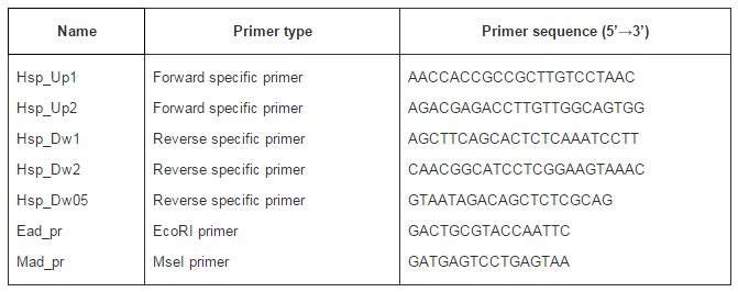
The combined approach proposed here has proved its usefulness for allowing the rapid cloning and characterization of specific conserved genes, as well for providing plant genomic fingerprinting information. An orthologous element for the Arabidopsis More Axillary Branching gene was successfully cloned and sequenced, for the first time, from several H. rosa-sinensis cultivars and from Hibiscus wild species. This general approach is particularly useful when dealing with plant species for which no or poor information is available at the genomic level. Minor changes could be made to the suggested protocol, i.e. the choice of the restriction enzyme could likely increase the efficiency of the method.

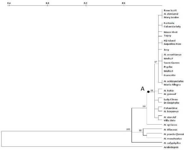
The high conservation sequence degree observed among commercial varieties and the Hibiscus species sexually compatible with H. rosa-sinensis are in agreement with the cytogenetic evidence produced by Singh and Khoshoo (1989), which showed that these inter-fertile species have contributed to the extensive genetic variability of H. rosa-sinensis. The revealed HibMAX2-like sequence analysis is consistent with secondary ranks of taxa (Sections) proposed by Pfeil and Crisp (2005) through chloroplast DNA analysis. In fact, the highest similarity values for the target sequence were achieved among the analyzed H. rosa-sinensis cultivars and the sexually compatible species, all belonging to the Lilibiscus Section, while the lower values were observed for species of different taxonomic Sections such as H. syriacus, H. panduriformis, H. moscheutos, H. tiliaceus and H. calyphyllus. Concerning the H. cannabinus, it could likely either do not possess the HibMAX2 gene, or possess a highly differentiated element, therefore not amplified. Kenaf is one of the fast-growing plants classified in the Furcaria Section of Hibiscus; it has both annual and biennial genotypes, often not branched. This differentiates from the other examined species (shrubs or small trees) characterized by perennial life cycle, with complex vegetative morphologies (Craven et al 2003). Moreover, a previous study (Braglia et al 2010) had revealed the lowest genetic similarity value between H. rosa- sinensis cultivars and kenaf defining this latter as the most distantly related species within the Hibiscus genus.

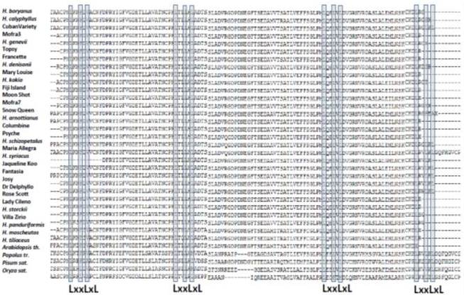
The occurrence of LRRs in the Hibiscus isolated fragment assigns this sequence among the F-box genes, one of the largest multigene superfamilies involved in shoot lateral branching growth. Members of this protein family function as subunits of the multiprotein Skp-Cullin-Fbox for polyubiquitination and degradation by the 26S proteasome (Xu et al 2009). In particular, the F-box LRR proteins confer substrate specificity to the SCF complex via their two distinct functional domains: the first domain (F-box) binds to another subunit of the SCF complex, the second domain (LRR repeats) interacts with specific proteins to be polyubitquinated (Stirnberg et al 2007).

Although the present results are the first step in the isolation of the whole Hibiscus specific element for MAX2 gene, the cloned fragment can be already investigated for association to the branched trait, to evaluate its utility in marker-assisted breeding schemes. Further studies including the isolation of a cDNA fragment (working back to the full length through RACE-PCR technique), the mRNA expression analysis and the functional variant identification are in turn necessary to better characterize the HibMAX2-like sequence, as well as to clarify its involvement in the axillary branch proliferation mechanisms.

Acknowledgements

We wish to thank Mr. Cesare Bianchini and Dr. Marco Ballardini for their support in managing the germplasm collection. Research founded by the Italian Ministry of Agriculture in the framework of the project “Risorse tecniche e genetiche per il florovivaismo (FLORIS)”.

References

Arite, T., Umehara, M., Ishikawa, S., Hanada, A., Maekawa, M., Yamaguchi, S. & Kyozuka, J. (2009). “D14, a Strigolactone-Insensitive Mutant of Rice, Shows an Accelerated Outgrowth of Tillers,” Plant & Cell Physiology, 50 1416—1424.
PublisherGoogle Scholar

Bennett, T., Sieberer, T., Willett, B., Booker, J., Luschnig, C. & Leyser, O. (2006). “The Arabidopsis MAX Pathway Controls Shoot Branching by Regulating Auxin Transport,” Current Biology, 16 553—563.
PublisherGoogle Scholar

Booker, J., Sieberer, T., Wright, W., Williamson, L., Willett, B., Stirnberg, P., Turnbull, C., Srinivasan, M., Goddard, P. & Leyser, O. (2005). “MAX1 Encodes a Cytochrome P450 Family Member that Acts Downstream of MAX3/4 to Produce a Carotenoid-Derived Branch-Inhibiting Hormone,” Developmental Cell, 8 443—449.
PublisherGoogle Scholar

Braglia, L., Bruna, S., Lanteri, S., Mercuri, A. & Portis, E. (2010) “An AFLP-based assessment of the genetic Diversity within Hibiscus rosa-sinensis and its Place within the Hibiscus Genus Complex,” Scientia Horticulturae, 123 (3) 372-378.
PublisherGoogle Scholar

Craven, L. A., Wilson, F. D. & Fryxell, P. A. (2003). “A Taxonomic Review of Hibiscus Sect. Furcaria (Malvaceae) in Western Australia and the Northern Territory,” Australian Systematic Botany, 16 (2) 185-218.
PublisherGoogle Scholar

Hillis, D. M. & Bull, J. J. (1993). “An Empirical Test of Bootstrapping as a Method for Assessing Confidence in Phylogenetic Analysis,” Systematic Biology, 42 182-192.
PublisherGoogle Scholar

Johnson, X., Brcich, T., Dun, E. A., Goussot, M., Haurogné, K., Beveridge, C. A. & Rameau, C. (2006). “Branching Genes are Conserved Across Species. Genes Controlling a Novel Signal in Pea are Co Regulated by Other Long-Distance Signals,” Plant Physiology, 142 1014—1026.
PublisherGoogle Scholar

Leyser, O. (2009). “The Control of Shoot Branching: An Example of Plant Information Processing,” Plant, Cell &Environment 32, 694—703.
PublisherGoogle Scholar

Liang, J., Zhao, L., Challis, R. & Leyser, O. (2010). “Strigolactone Regulation of Shoot Branching in Chrysanthemum (Dendranthema Grandiflorum),” Journal of Experimental Botany 61(11), 3069-3078.
PublisherGoogle Scholar

Lumba, S., Cutler, S. & McCourt, P. (2010). “Plant Nuclear Hormone Receptors: A Role for Small Molecules in Protein-Protein Interactions,” Annual Review of Cell and Developmental Biology 26, 445—69.
PublisherGoogle Scholar

McSteen, P. & Leyser, O. (2005). “Shoot Branching,” Annual Review of Plant Biology 56, 353—374.
PublisherGoogle Scholar

Morris, S. E., Turnbull, C. G. N., Murfet, I. C. & Beveridge, C. A. (2001). “Mutational Analysis of Branching in Pea. Evidence that Rms1 and Rms5 Regulate the Same Novel Signal,” Plant Physiology 126, 1205-1213.
PublisherGoogle Scholar

Pfeil, B. E. & Crisp, M. D. (2005). “What to Do with Hibiscus? A Proposed Nomenclatural Resolution for a Large and Well Known Genus of Malvaceae and Comments on Paraphyly,” Australian Systematic Botany 18(1), 49-60.
PublisherGoogle Scholar

Singh, F. & Khoshoo, T. N. (1989). ‘Cytogenetic Basis of Evolution in Garden Hibiscus,’ Nucleus 32, 62—67.
Google Scholar

Snowden, K. C. & Napoli, C. A. (2003). “A Quantitative Study of Lateral Branching in Petunia,” Functional Plant Biology30, 987-994.
PublisherGoogle Scholar

Stirnberg, P., Furner, I. J. & Leyser, H. M. O. (2007). “MAX2 Participates in an SCF Complex which Acts Locally at the Node to Suppress Shoot Branching,” The Plant Journal 50, 80—94.
PublisherGoogle Scholar

Stirnberg, P., van den Sande, K. & Leyser, H. M. O. (2002). “MAX1 and MAX2 Control Shoot Lateral Branching in Arabidopsis,” Development 129, 1131—1141.
PublisherGoogle Scholar

Van de Peer, Y. & De Wachter, R. (1994). “Treecon for Windows: A Software Package for the Construction and Drawing of Evolutionary Trees for the Microsoft Windows Environment,” Computer Applications in the Biosciences 10, 569—570.
PublisherGoogle Scholar

Vos, P., Hogers, R., Bleeker, M., Reijans, M., van de Lee, T., Homes, M., Freiters, A., Pot, J., Peleman, J., Kuiper, M. & Zabeau, M. (1995). “AFLP: A New Technique for DNA Fingerprinting,” Nucleic Acids Research 23, 4407-4414.
PublisherGoogle Scholar

Wang, Y., Sun, S., Zhu, W., Jia, K., Yang, H. & Wang, X. (2003). ‘Strigolactone/MAX2-Induced Degradation of Brassinosteroid Branscriptional Effector BES1 Regulates Shoot Branching Developmental,’ Cell 27, 681—688.

Xu, G., Ma, H., Nei, M. & Kong, H. (2009). “Evolution of F-Box Genes in Plants: Different Modes of Sequence Divergence and Their Relationships with Functional Diversification,” Proceedings of the National Academy of Sciencesof the United States of America 106, 835—840.
PublisherGoogle Scholar

Shares