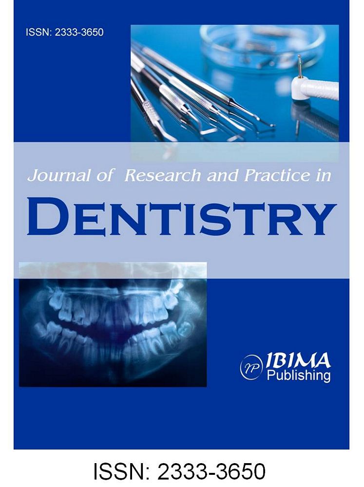
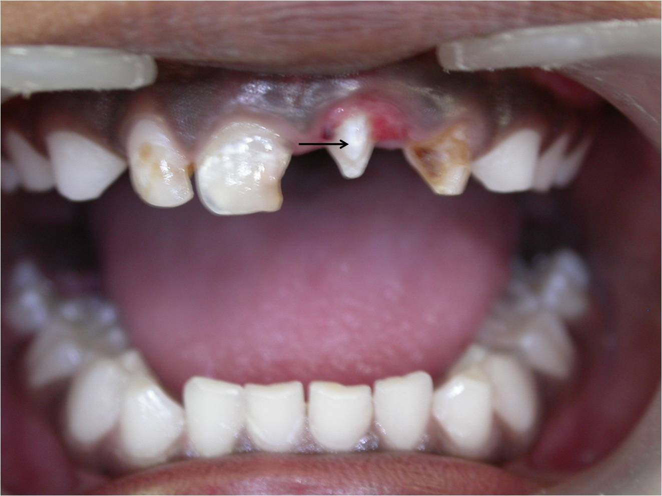
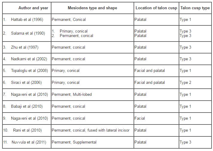
The majority of times, talon cusp is seen on lingual surface of anterior teeth. Literature documentation of facial talons in a supernumerary tooth is very few and little or no epidemiological information is available. Extensive literature search revealed only three publications of facial talon in supernumerary tooth (Siraci et al 2006, Topaloglu et al 2008, Nagaveni et al 2010). One publication is by Siraci et al (2006), who reported both facial and palatal talons on a supernumerary primary incisor with pulp extensions detected by a micro-CT. Another case is reported by Topaloglu et al (2008), who also found both facial and palatal talons in primary mesiodens. Recently Nagaveni et al (2010) in their epidemiological survey of mesiodens occurring in 2500 Davangere (India) children reported a case of facial talon in maxillary mesiodens tooth. Therefore, the development of talon on the facial aspect of a supernumerary tooth further constitutes a rarer entity. In all our cases, talon cusps were found on the facial surface of mesiodens teeth thereby being an extremely rare occurrence.
Jowharji et al (1992) commented on facial talon anomaly and suggested that an alteration to the conventional definition of talon cusps be made; taking into consideration that talon cusp may be found on the facial aspect of teeth. However, they (Jowharji et al 1992) limited the definition to incisor teeth. Later McNamara et al (1997) reported a facial talon on maxillary canine tooth in 1997, and suggested that the definition should include teeth in addition to the incisor group and be extended to include the facial aspect too. According to these authors’ opinion and from our cases, we strongly recommend, for a better understanding of this condition, that the definition should be further extended to include the mesiodens tooth.
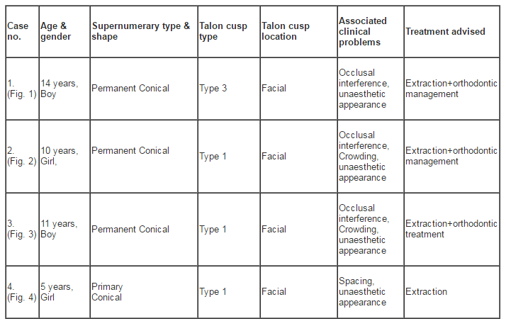
Hattab et al (1996) have classified the talon cusps into three types based on the degree of formation and extension. Type 1 talon refers to a morphologically well-delineated additional cusp that prominently projects from the palatal surface of a primary or permanent anterior tooth, and extends at least half the distance from the CEJ to the incisal edge. Type 2 talon is an additional cusp of a millimeter or more, extending less than half the distance from the CEJ to the incisal edge. It may blend with the palatal surface or stand away from the rest of the crown. And type 3 is enlarged or prominent cingula and their variations, i.e., conical, bifid, or tubercle like. The same classification can be employed in classifying the facial talon cusps too. Therefore, based on this classification, our reported cases were categorized as type 3 Talon cusp in first case as the talon appeared like a small tubercle. In second, third and fourth case, the talon extended more than half distance from the CEJ to the incisal edge. Hence they were classified as type 1 talon.
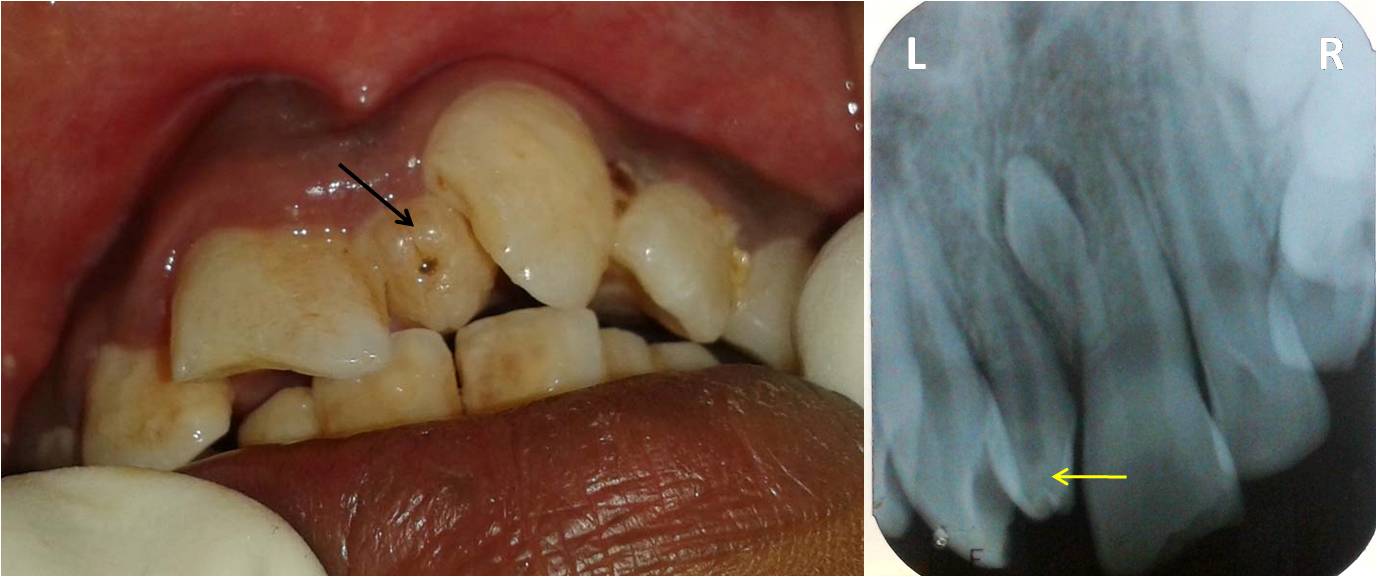
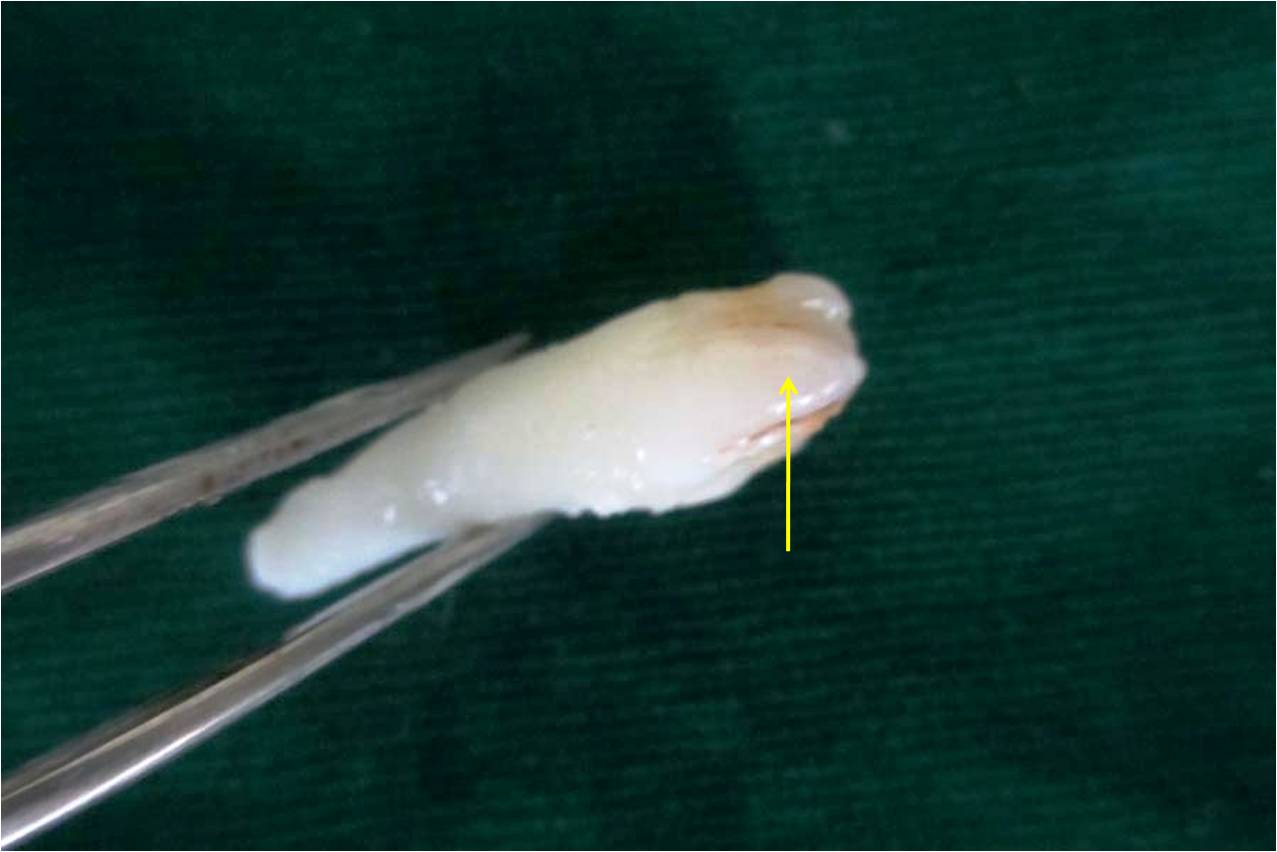
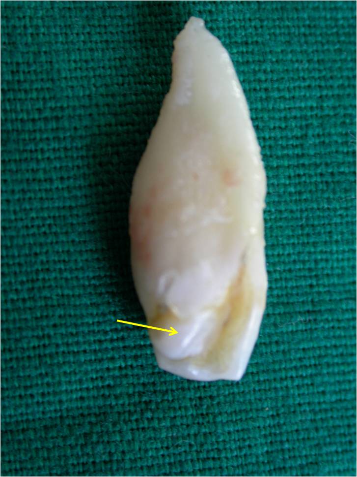
Supernumerary teeth in the primary dentition are usually normal or conical in shape. In the permanent dentition, they show great variation in morphology. Primosch (1981) classified them into supplemental or eumorphic and rudimentary or dysmorphic types (conical, tuberculate and molariform), according to their shapes. According to Koch et al in (1986), supernumerary teeth may be classified as 56% conical, 12% tuberculate, 11% supplemental, and 12% other configurations. Recently, Nagaveni et al (2010) presented a new form of mesiodens (multi-lobed mesiodens) associated with a palatal talon. In the presented four cases, the mesiodens tooth was classified as conical type based on the above classification.
Treatment modality for talon cusps may differ depending on each case and presenting complaints (Hattab et al 1996). In this report, extraction of involved tooth was advised in all three cases, as the affected teeth were a mesiodens which caused occlusal interference and esthetic problems followed by orthodontic treatment.
1. Babaji P, Sanadi F, and Melkundi M. (2010) “Unusual case of a talon cusp on a supernumerary tooth in association with a mesiodens,” Journal of Dental Research Dental Clinics Dental Prospects, 4(2):60-3.
Google Scholar
2. Buanviaje T. M, and Rapp R. (1984) “Dental anomalies in children: A clinical and radiographic survey,” Journal of Dentistry for Children, 51:42-6.
3. Chawla H. S, Tewari A, and Gopalakrishnan N. S. (1983) “Talon cusp — a prevalence study,” Journal of Indian Society of Pedodontics and Preventive Dentistry, 28-34.
4. Chen R. J, and Chen H. S. (1986) “Talon cusp in primary dentition,” Oral Surgery, Oral Medine Oral Pathology Oral Radiology Endodontics, 62: 67-72.
Publisher – Google Scholar
5. Davis P. J, and Brook A. J. (1985) “The presentation of talon cusp: Diagnosis, clinical features, associations and possible etiology,” British Dental Journal, 159: 84-88.
6. de Saousa S. M. G, Tavano S. M. R, and Bramante C. M. (1999) “Unusual case of bilateral talon cusp associated with dens invaginatus,” International Endodontic Journal, 32: 494-8.
Publisher – Google Scholar
7. Hattab F. N, Yassin O. M, and Al-Nimri K. S. (1990) “Talon cusp in permanent dentition associated with other dental anomalies: Review of literature and report of seven cases,” Journal of Dentistry for Children, 63: 368-76.
8. Hattab F. N, Yassin O. M, and Al-Nimri K. S. (1995) “Talon cusp — clinical significance and management: case reports,” Quintessence International, 26:115-20.
9. Henderson H. Z. (1977) “Talon cusp: a primary or permanent dentition anomaly,” Journal of Indian Dental Association, 56: 45-56.
10. Jowharji N, Noonan R. G, and Tylka J. A. (1992) “An unusual case of dental anomaly: A facial talon cusp,” ASDC J Dent Child , 59(2): 156-158.
11. Koch H, Schwartz O, and Klausen B. (1986)“Indications for surgical removal of supernumerary teeth in the premaxilla,” International Journal of Maxillofacial Surgery, 15: 273-281.
Publisher – Google Scholar
12. Mader C. L. (1981) “Mandibular talon cusp,” Journal of American Dental Association, 103: 244-246.
13. McNamara T, Haeussler A, and Keane J. (1997) “Facial talon cusps,” International Journal of Pediatric Dentisty, 7: 259-262.
Publisher – Google Scholar
14. Nadkarni U. M, Munshi A, and Damle S. G. (2002) “Unusual presentation of talon cusp: two case reports,” International Journal of Pediatric Dentistry, 12: 332-335.
Publisher – Google Scholar
15. Nagaveni N. B, Umashankara K. V, Sreedevi, Reddy B. P, Radhika N. B, and Satisha T. S. (2010) “Multi-lobed mesiodens with a palatal talon cusp — A rare case report,” Brazilian Dental Journal, 21(4): 375-378.
Publisher – Google Scholar
16. Nagaveni NB, Sreedevi B, Praveen BS, Praveen Reddy B, Vidyullatha BG, and Umashankara KV. (2010) “Survey of mesiodens and its characteristics in 2500 children of Davangere city, India,” European Journal of Paediatric Dentistry, 11(4): 185-188.
Google Scholar
17. Nuvvula S, Pavuluri C, Mohapatra A, and Nirmala S.V. (2011) “Atypical presentation of bilateral supplemental maxillary central incisors with unusual talon cusp,” Journal of Indian Society of Pedodontics and Preventive Dentistry,” 29(2):149-54.
Publisher – Google Scholar
18. Primosch R. E. (1981) “Anterior supernumerary teeth. Assessment and surgical intervention in children,” Pediatric Dentistty, 3: 204-215.
19. Rani A. K, Metgud S, Yakub S. S, Pai U, Toshniwal N. G, and Bawaskar N. (2010) “Endodontic and esthetic management of maxillary lateral incisor fused to a supernumerary tooth associated with a talon cusp by using spiral computed tomography as a diagnostic aid: a case report,” Journal of Endodontics, 36: 345-349.
Publisher – Google Scholar
20. Salama F. S, Hanes C. M, Hanes P. J, and Ready M. A. (1990) “Talon cusp: a review and two case reports on supernumerary primary and permanent teeth,” Journal of Dentistry for Children, 57: 147-149.
21. Sedano H. O, Freyre I. C, and de La Garza M. L. (1989) “Clinical orodental abnormalities in Mexican children,” Oral Surgery Oral Medicine Oral Pathology Oral Radiology Endodontics, 68:300-11.
Publisher – Google Scholar
22. Siraci E, Gungor H. C, Taner B, and Cehreli Z. C. (2006) “Buccal and palatal talon cusps with pulp extensions on a supernumerary primary tooth,” Dentomaxillofacial Radiology, 35: 469-472.
Publisher – Google Scholar
23. Topaloglu A. K A, Eden E, Ertugrul F, and Sutekin E. (2008) “Supernumerary primary tooth with facial and palatal talon cusps: A case report,” Journal of Dentistry for Children, 75: 309-312.
24. Zhu J. F, King D. L, and Henry R. J (1997). “Talon cusp associated with adjacent supernumerary tooth,” General Dentistry, 45(2):178-81.