Introduction
Foot and mouth disease (FMD) is a highly contagious disease of cloven footed animals. It is economically the most important disease of livestock worldwide (Bachrach, 1968; Kitching 1992). This is a highly transmissible notifiable viral disease (Nunez et al., 1998). Despite low mortality rates, FMD severely decreases livestock production and leads to trade restrictions on animals and livestock products.
The FMD virus (FMDV; genus Aphthovirus; family Picornaviridae) consists of seven serotypes (O, A, C, Asia1, SAT-1, SAT-2 & SAT-3) (Domingo et al, 2003). The serotypes O and A are currently endemic worldwide, Asia 1 is currently prevalent in Asia including India, and the serotype C has not been reported from India since 1995 (Shah et al., 2011) and worldwide since 2004. The vaccine against FMD currently manufactured in the country is a trivalent vaccine consisting of serotypes O, A and Asia 1 (Hemadri et al., 2008).
Accurate diagnosis of the infection with FMDV is of prime importance for the control and eradication of the disease in endemic areas. Currently, there is no universal FMD vaccine (Aftosa, 2007). Vaccination with one serotype does not protect the animal against other serotypes. Identification of the serotype is therefore essential as the vaccines must closely match the serotype of the infecting strain. Although several sensitive diagnostic tests like antigen ELISA, Complement Fixation Test (CFT) and Reverse Transcription Polymerase Chain Reaction (RT PCR) are available to confirm the serotype or genotype of the circulating FMDV, none of them is applicable in field conditions in developing countries due to their cumbersome nature, high costs and requirement of skilled personnel and well equipped laboratories. A simple and cheap pen side kit for identifying the serotype of the virus responsible for infection in an area can be useful in vaccination against prevalent serotypes and avoiding vaccination against serotypes not prevalent in the area. Such a test can be conducted by vets with minimum instructions. It would save time and resources and also circumvent the problems associated with the transportation of samples to the lab at low temperature.
Material and Methods
Blood samples of cattle and buffaloes vaccinated with the trivalent FMD vaccine (Indian Immunologicals; strains O IND R2/75, A IND 40/00, and Asia 1 IND 63/72) were obtained from an organized dairy farm of Ludhiana and private farms of Sangroor, Ropar and Kapurthala districts of Punjab state. Serum was separated from these blood samples and 0.05% Merthiolate was added in it to preserve the samples. The sera were kept at -80oC before testing. All reagents were equilibrated at room temperature before use.
Preparation of Latex Beads for Agglutination: The latex beads available commercially were procured from Sigma, USA. The beads (300µl) were suspended in 15 ml of 0.05 M Glycine Saline and centrifuged at 12,500g for 15 minutes. The above procedure was repeated to collect the beads.
Coating of Antigen on to Latex Beads: The FMD virus of O, A, Asia 1 serotypes were coated on to the Latex beads as per the protocol of Hay et al. (2002). The following vaccine strains of FMD virus were used for coating onto latex beads: strains O IND R2/75, A IND 40/00, and Asia 1 IND 63/72. Inactivated viruses of all the three serotypes were individually mixed with 10% (w/v) latex beads suspension prepared as above in the final volume of 2.5 ml Glycine Saline to achieve a final protein concentration of 10mg/ml. Incubation at 4oC for 8h was provided to achieve latex protein binding. Later, the suspension was centrifuged at 12,500g for 15 minutes to collect the beads. The beads were washed again with 2.5 ml of Glycine Saline (0.05 M) and were collected by centrifugation at 12,500g for 15 minutes. Finally, the beads were suspended in 2.5 ml of 0.25M Glycine Saline and stored at 4oC for further use.
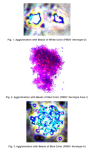
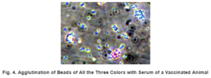
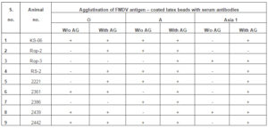
Coating onto Colored Latex Beads: FMD virus of the three serotypes i.e. O, A, Asia 1 were used for the coating of the inactivated virus on to different colored (Red, Blue and White) latex beads as per the protocol of Hay et al. (2002). The antigens (FMDV O, A and Asia 1), were coated on white, blue and red latex beads, respectively, as per the protocol stated above.
Latex Agglutination Test: All the serum samples were tested against all the three differentl colored latex beads coated with the viruses of different serotypes. Equal quantity (10µl) of serum sample and inactivated virus coated on to the latex beads was put on clean, grease – free glass slide. The serum sample and the latex beads were mixed thoroughly using a clean wooden toothpick and observed for clump formation indicative of agglutination within one minute. The samples were classified as negative, weak positive, positive and strong positive depending on the intensity of the agglutination.
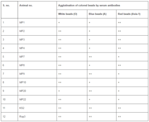
Agglutination with Colored Latex Beads: All the serum samples were tested against all the three differently colored latex beads. Ten microlitres of serum sample and ten microlitres of either of the three antigens coated on to different latex beads was put on a clean grease- free glass slide. The serum sample and the latex beads were mixed thoroughly using a clean wooden toothpick and observed for the clump formation of various colors indicative of agglutination with a particular serotype of FMDV.
LPB ELISA: Titers of antibodies against FMDV O, A and Asia-1 in sera from 6 FMD vaccinated animals were assessed at the Regional Disease Diagnosis Laboratory (RDDL), Jalandhar employing the Liquid Phase Blocking ELISA (LPB ELISA) as per the OIE protocol (Hamblin et al, 1986).
Results
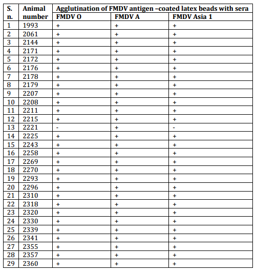
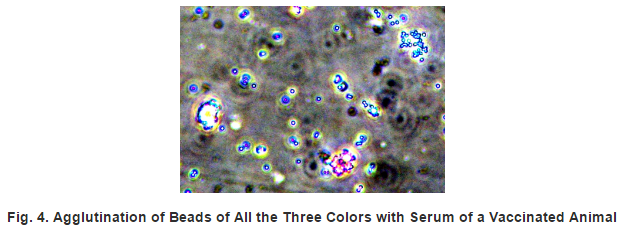
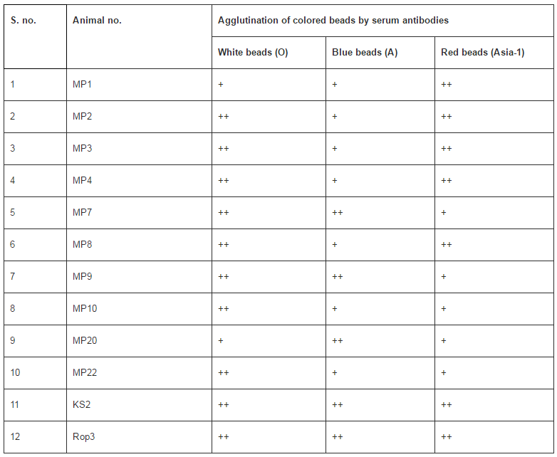
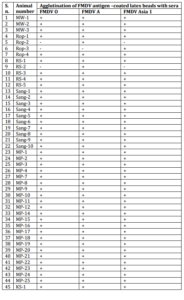
Detection of Antibodies to Different Serotypes of FMD Virus by the Pen Side Kit: The prototype of an agglutination based new pen side kit for antibody detection in serum to differentiate infection with different serotypes was developed by conjugating inactivated FMDV of serotypes O, A and Asia-1 with latex beads/microspheres of different colors. The kit was used for detection of serotype specific antibodies in serum samples from 142 animals vaccinated with polyvalent vaccine for FMD Virus O, A & Asia-1. Serotype — specific antibodies could be detected against all the three serotypes in the sera of 93.66% of the animals (Tables 1 & 2; Figs. 1 to 4).
Table 1: Agglutination of Individual FMDV Antigen—Coated Single Color Beads with Sera of Vaccinated Animals from the Dairy Farm


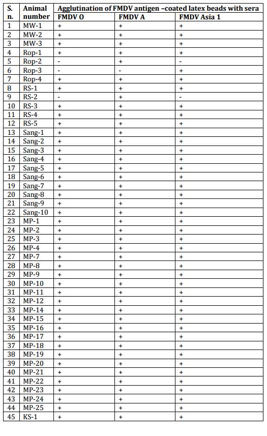
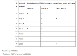
Table 2: Agglutination of FMDV-Coated Single Color Latex Beads with Sera of Field Animals
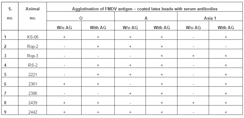
However, antibodies against A serotype could not be detected in the serum of 1 (0.7%) animal, and against Asia-1 serotype in the sera of 2 (1.40%) animals, respectively. Four (2.80%) serum samples were double negative for O and Asia-1 serotypes and one (0.7%) sample each was double negative for O and A as well as A and Asia-1 serotypes, respectively. Antiglobulin was used to enhance the sensitivity of the kit through cross-linking of smaller clumps. Detection of antibodies to FMDV A was then possible in the sera of all the three previously negative animals. However, 2 out of 5 and 1 out of 7 serum samples were still negative for antibodies to FMD Virus O and Asia-1, respectively (Table 3).
Table 3: Antiglobulin Facilitated Detection of Agglutination with False Negative Sera
It may be possible that these animals were exposed to the FMDV serotype A only and could generate immune response to this serotype alone. All the remaining sera which were previously negative gave positive reaction on addition of antiglobulin. The multiplex agglutination of sera from vaccinated animals could detect antibodies to all the three serotypes of FMDV in the same serum sample simultaneously (Table 4).
Table 4. Testing of Sera with Multiplex Agglutination for Three Serotypes of FMDV
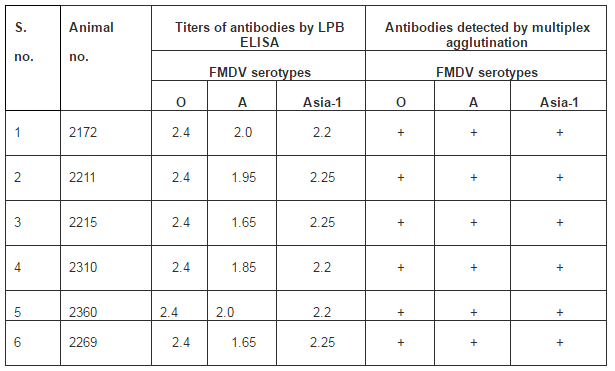
The results were verified by determining the antibody titers to the virus by LPB ELISA of sera from six vaccinated animals (Table 5).
Table 5: A Comparison of Antibodies to FMDV Serotypes in Vaccinated Animals Detected by Multiplex Agglutination and LPB ELISA

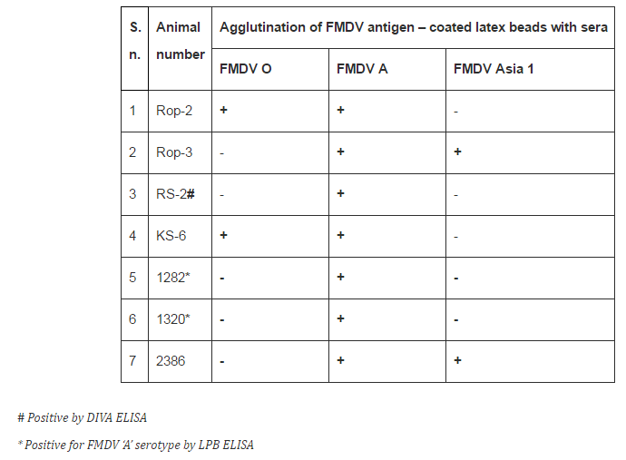
The detection of antibodies for a single serotype A by the multiplex agglutination was confirmed by LPB ELISA (Table 6).
Table 6: Antibodies to Single or Two Serotypes of FMDV in Sera of Animals Detected by the Agglutination Kit
Since monovalent vaccines were not available commercially, monospecific sera raised in rabbits were used to ascertain the serotype specificity of the antibodies detected by the FMDV conjugated beads.
Discussion:
It has been reported earlier that about 85% of the outbreaks of FMD in India are caused by serotype O, 8-10% by A and rest due to Asia 1 (Biswal et al., 2012) indicating that protection against serotype O is very important from the point of view of FMD control.
Multiplex immunoassays offer the capacity to detect multiple analytes in a single assay, thereby reducing hands-on time, specimen volume requirements, and turnaround time. These advantages have been demonstrated in several recent studies evaluating MFI assays for the detection of autoantibodies in autoimmune disorders, as well as in the evaluation of Epstein-Barr and herpes simplex virus infections (Binnicker et al., 2008, 2010; Hanly, et al, 2010; Kaul et al, 2009). Gomez et al (2010) evaluated the performance of a novel multiplex flow immunoassay for the detection of antitreponemal IgG- and IgM-class antibodies. The multiplex assay showed serological agreement with EIA.
We report here the proof of concept of a new agglutination based pen side kit for antibody detection in serum to differentiate infection with different serotypes of FMDV employing latex beads/microspheres of different colors. The prototype kit was used for detection of FMD virus serotype specific antibodies in serum samples from buffaloes vaccinated with a polyvalent vaccine for FMD Virus O, A and Asia-1. Detection of antibodies to FMDV A was possible in the sera of all the three false negative animals with the aid of antiglobulin. The new kit holds the promise of a candidate penside kit for large scale low-cost screening of animals in the FMD — endemic areas to identify the individual serotype prevalent in the area. This may also be adapted with suitable minor modifications as a DIVA kit to differentiate animals vaccinated with the polyvalent vaccine from those infected with only one or two serotypes of FMDV or with other exotic serotypes not included in the available vaccines (like SAT-1, SAT-2 or SAT-3 or C). Thus, detection of antibody by multiplex agglutination employing antigen coated beads – different colored beads for different serotypes, can be used as a pen side kit for differential diagnosis where antiglobulin may impart greater sensitivity and may be helpful in discriminating real negative from the false negative serum samples. The prototype assay may be further refined by using synthetic peptide antigens of viruses instead of inactivated whole virus.
Acknowledgements:
We are thankful to Dr. Ravindra Sharma, LLRUVAS, Hisar, India and Dr. Ashwini Kumar, RDDL, Jalandhar, India for help.
References:
Aftosa, F. (2007). “Foot and Mouth Disease,” The Centre for food Security and Public Health, Ames, USA. 1-6.
Publisher
Bachrach, H. L. (1968). “Foot-and-Mouth Disease Virus,” Ann Rev Microbiol. 22: 201.
Publisher – Google Scholar
Binnicker, M. J., Jespersen, D. J. & Harring, J. A. (2010). “Evaluation of Three Multiplex Flow Immunoassays Compared to an Enzyme Immunoassay for the Detection and Differentiation of Igg Class Antibodies to Herpes Simplex Virus Types 1 and 2,” Clin. Vaccine Immunol. 17: 253-257.
Publisher – Google Scholar
Binnicker, M. J., Jespersen, D. J., Harring, J. A., Rollins, L. O. & Beito, E. M. (2008). “Evaluation of a Multiplex Flow Immunoassay for Detection of Epstein-Barr virus-Specific Antibodies,” Clin. Vaccine Immunol. 15: 1410-1413.
Publisher – Google Scholar
Biswal, J. K., Sanyal, A., Rodriguez, L. L., Subramaniam, S., Arzt, J., Sharma, G. K., Hammond, J. M., Parida, S., Mohapatra, J., Mathapati, B. S., Dash, B. B., Ranjan, R., Rout, M., Venketaramanan, R., Misri, J., Krishna, L., Prasad, G., Pathak, K. M. L. & Pattnaik, B. (2012). “Foot-and-Mouth Disease: Global Status and Indian Perspective,” Indian J Anim Sci 82: 109-131.
Publisher – Google Scholar
Domingo, E., Escarmis, C., Baronowski, E., Ruiz-Jarabo, C. M., Carrillo, E., Núñez, J. I. & Sobrino, F. (2003). “Evolution of Foot and Mouth Disease Virus,” Virus. 9147-63.
Publisher – Google Scholar
Gomez, E., Jespersen, D. J. , Harring, J. A. & Binnicker, M. J. (2010). “Evaluation of the Bio-Rad BioPlex 2200 Syphilis Multiplex Flow Immunoassay for the Detection of IgM- and IgG-Class Antitreponemal Antibodies,” Clin Vaccine Immunol. 17(6): 966—968.
Publisher – Google Scholar
Hanly, J. G., Thompson, K., McCurdy, G., Fougere, L., Theriault, C. & Wilton, K. (2010). “Measurement of Autoantibodies Using Multiplex Methodology in Patients with Systemic Lupus Erythematosus,” J. Immunol. Methods 352: 147-152.
Publisher – Google Scholar
Hay, F. C., Westwood, O. M. R., Nelson, P. N. & Hudson, L. (2002). “Antibody Interactions with Antigens,” Chapter 3. Practical Immunology 4th edn. 71-114.
Publisher
Hemadri, D., Sanyal, A., Tamil Selvan, R. P., Saravanan, S., Mohapatra, J. K., Lalkrishna, Bujarbaruah, K. M. & Pattnaik, B. (2008). “Constraints and Opportunities for the Control of Foot and Mouth Disease in India,” presented at The Global Control of FMD – Tools, Ideas and Ideals — Erice, Italy.
Publisher
Kaul, R., Johnson, K., Scholz, H. & Marr, G. (2009). “Performance of the BioPlex 2200 Autoimmune Vasculitis Kit,” Autoimmun Rev. 8: 224-227.
Publisher – Google Scholar
Kitching, R. P. (1992). “Foot-and-Mouth Disease: Current World Situation,” Vaccine 17: 1772.
Publisher – Google Scholar
Nunez, J. I., Blanco, E., Hernandez, T., Gomez, T. C., Martin, M. J., Dopazo, J. & Sobrino, F. (1998). “A RT PCR Assay for the Differential Detection of Viral Vesicular Diseases in Swine,” J Virol Meth, 72: 227-235.
Publisher – Google Scholar
Shah, K. A., Jan, F. A., Andrabi, A. & Kaloo, F. A. (2011). “Incidence, Serotyping and Epidemiology of Foot and Mouth Disease in Kashmir,” Online Vet J. 6: 1.
Publisher