Volume 2026,
Article ID 526985,
Communications of the IBIMA,
16 pages,
DOI: doi.org/10.5171/2026.526985
Received date: 3 June 2026; Accepted date: 11 June 2026; Published date: 21 July 2026
Academic Editor: Srinivasulu Battu
Cite this Article as:
Ahmed A. Alani, Jyoti Pareek and Vishal Prajapati (2026)," A Review of Using Machine Learning Techniques for Skin Disease Prediction", Communications of the IBIMA, Vol. 2026 (2026), Article ID 526985, https://doi.org/10.5171/2026.526985
One of the primary problems in our society is skin diseases, due to their severe effects on both the body and the mental health of individuals. Early detection and diagnosis of these diseases can have a significant impact on the success of treatments. The skills and experience of specialists play a crucial role in determining effective methods for diagnosing and treating skin lesions. In the early stages, advanced techniques were developed to automatically test skin for diseases. In recent years, skin diseases have increasingly been diagnosed with the help of artificial intelligence through machine learning algorithms, which are trained on vast amounts of data already available in the healthcare sector. In this research report, we comprehensively investigated previous studies on the use of machine learning in skin disease classification. Skin diseases were successfully classified in several studies with varying levels of diagnostic accuracy. Some studies used image processing and feature extraction for this purpose, while others focused on specific types of skin diseases by utilizing clinical features obtained from tissue analyses of the affected areas. This research report concludes that using image processing methods, accuracy ranged between 50% and 100%. Another method, involving tissue analysis, achieved an excellent accuracy rate of 94% or higher. The findings present a review of the related studies conducted in the literature and focus on the recent research gaps.
In general, human skin functions as a protective barrier against outside elements, serving as a buffer between the environment and the body. Various types and variations of skin diseases have different causes, inclusive of internal ones associated with hormones and body glands such
as pimples or outside associated with air pollution or exposure to sunlight along with rashes. Skin conditions often result from internal irregularities, infections, pollution, genetic issues, exposure to ultraviolet (UV) light, and other factors. Current reports indicate that over 2000 different types of skin diseases are prevalent, affecting over 20% of global individuals at certain times in their lives [1]. Worldwide statistics show that skin illnesses cause 1.79% of human physical limitations worldwide [2]. About 30% to 70% of individuals suffer from skin-related issues in different parts of the world [3]. Skin diseases can manifest across all demographics and seasons, making diagnosis a complex task. Combining medical and technological expertise is crucial to effectively treating these conditions. Technological advancements, particularly artificial intelligence (AI), can aid in early detection and prompt treatment [4]. Researchers are currently developing AI-based models to assist dermatologists in accurately diagnosing skin diseases, minimizing the risk of misdiagnosis. [5] This survey aims to examine the latest skin disease classification and prognosis using Machine Learning Techniques. It entails an in-depth review of a recent model for skin disease diagnosis, detailing the machine-learning algorithm and the associated dataset. Additionally, the review provides an assessment of the predictive capabilities of each model in diagnosing various skin conditions. The subsequent section delves into Skin Diseases: Types, Prevalence, and Impact, Machine Learning Techniques to classify Skin Diseases, datasets, techniques for Feature Extraction and Selection, Model Development and Evaluation, Challenges, and Limitations, while the last Section discusses the Future Directions and Research Opportunities.
Literature Review
Early-stage Image-processing and Detection Diseases
Image processing has been used by several scientists to identify the type of disease that might affect the skin. In 2012, color images were utilized to classify skin diseases without the involvement of doctors. The proposed system involved two different detection stages: the first stage used color image processing to identify the affected skin through k-means clustering and color gradient techniques, while the second stage applied an artificial neural network to identify the kind of skin disease. Six different skin conditions were investigated, and the proposed system achieved accuracies of 95.99% and 94.016% for the first and second stages, respectively. In 2014, skin diseases were detected through feature extraction from skin images, with the accuracy dependent on the number of features extracted. Their method reached up to 90% accuracy for nine different skin disease types. In 2015, researchers applied various image processing segmentation algorithms to detect melanoma in its early stages [6], focusing on extracting features from the edges and boundaries of affected skin areas. In the same year, the studies proposed a method to detecting diseases in dark skin using a specialized algorithm on images from various diseases sources [7].
Transition to Combined Computer Vision with Machine Learning
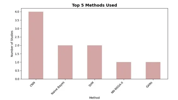
In 2016, researchers combined computer vision with machine learning as a new method to diagnose skin diseases. In this approach, the features in the images were extracted using computer vision, while the skin diseases were classified using machine learning. The authors reported 95% accuracy in detecting the diseases. In this year, studies introduced a support vector machine (SVM) method to classify skin diseases such as basal cell carcinoma (BCC), seborrheic keratosis (SK), nevus, and melanoma, achieving higher accuracy than other methods [8]. Another team developed a system to automatically detect eczema and assess its severity using segmentation, feature extraction (color, texture, borders), and SVM classification. Another type of disease known as chronic skin disease can spread rapidly to different areas of the affected body, causing severe consequences. Several scientists have devoted their efforts to detecting such diseases. For example, in 2016, several researchers developed a method to automatically detect eczema and define its severity using a computer system. The detection method involved three steps. In the first step, the affected skin was detected based on segmentation. In the second step, a set of features such as color, texture, and borders were extracted. Finally, in the last step, SVM was used to determine the severity of eczema. See Fig.1. show the popular methods and their frequency used for development
Figure 1: Bar graph for popular methods and their frequency used for development
Advanced: Fusion-level Deep Learning Models
The studies and researcher shifted toward deep learning approaches. One study proposed a CNN-based model to diagnose melanoma in its early stages, The proposed model classifies three types of melanomas: malignant melanoma, superficial spreading melanoma, and nodular melanoma. The CNN model was evaluated using data retrieved from the website dermnetnz.org. Experimental results show that the proposed model achieved a diagnosis accuracy of 88.83%[9]. An ensemble meta-strategy for the identification of ESD was provided by the study [10] and includes psoriasis, lichen planus, seborrheic dermatitis, pityriasis rosea, chronic dermatitis, and pityriasis rubra pilaris. The study examined many algorithms for classifying ESD based on clinical and histological data, including RF, DT, NB, KNN, and Multi-Layer Perceptron. The preprocessing techniques applied to the dataset included data cleaning, feature selection, data transformation, data splitting, and cross-validation. The model obtained an accuracy of 97.8% when assessed using accuracy, precision, recall, and F-1 score.
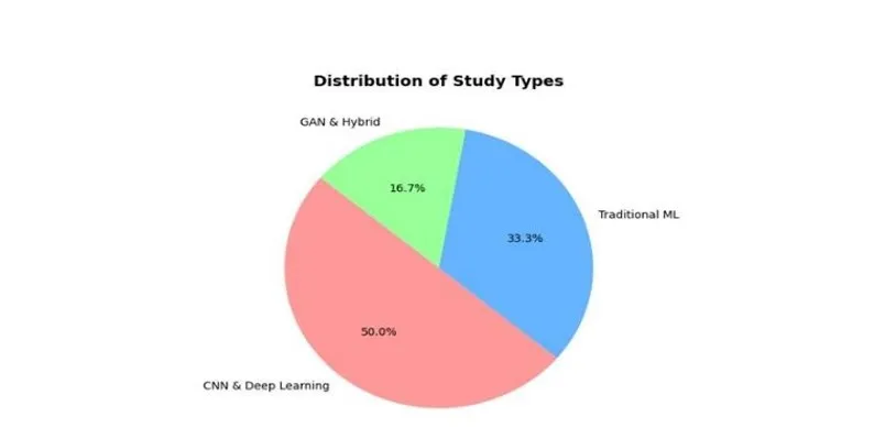
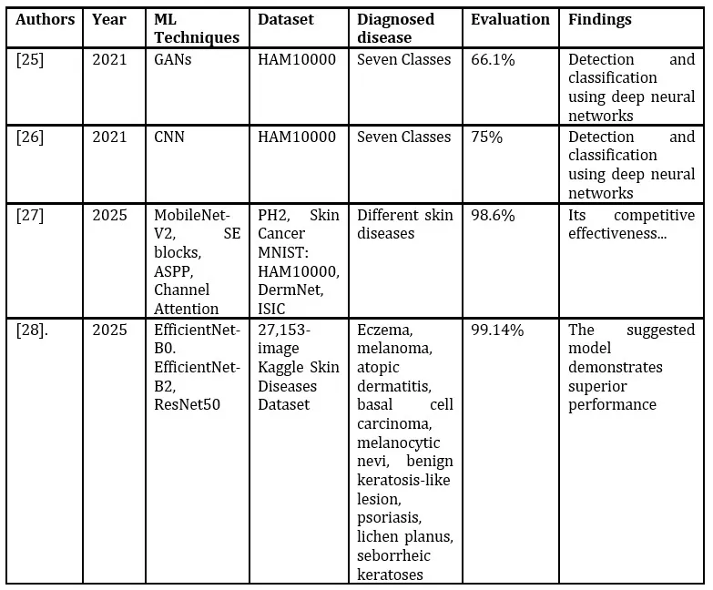
Additionally, to improve performance, the author [11] introduced a novel skin disease categorization model that makes use of cutting-edge deep learning. The MobileNet-V2 architecture incorporates squeeze-and-excitation networks, atrous spatial pyramid pooling, and the channel attention mechanism. Diverse datasets, including PH2, Skin Cancer MNIST: HAM10000, DermNet, and Skin Cancer ISIC, are utilized; scaling and mean subtraction normalization are implemented. The MobileNet-V2 backbone captures hierarchical features, whereas ASPP integrates multi-scale contextual information to provide a feature map. A study on self-attention techniques enhances the extraction of inter-channel linkages and contextual information, hence improving feature discrimination. The model achieves an accuracy of 98.6%. [12] developed a fusion-level deep learning model that enhances reliability and the classification effectiveness for skin diseases. The model architecture integrates three convolutional neural networks (CNNs): EfficientNet-B0, EfficientNet-B2, and ResNet50, which function independently within separate branches. The fusion mechanism concludes its function by sending extracted data to dense and dropout layers for generalization and dimensionality reduction. This research employed analyses of the 27,153-image Kaggle Skin Diseases Image Dataset. The assessment of the suggested model demonstrated an accuracy of 99.14%.see Fig.2 show Distribution of approaches for skin diseases detection field
Figure 2: Distribution of approaches for skin diseases detection field so far
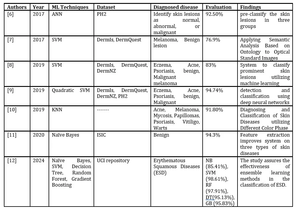
Table1: Machine learning based approaches and research
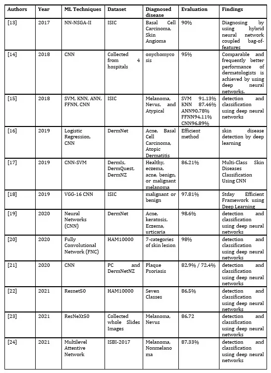
Table2: Deep learning-based approaches and research
Table3 Advanced Deep learning approaches
Skin Diseases: Overview and Importance of Detection
Overview of Skin Diseases
Skin diseases are common health problems and account for a significant portion of visits to healthcare facilities. They are of different types, and each disease has its own impact factor. Skin diseases can be acute, chronic, and in some cases lifelong or even life-threatening if not treated or recognized properly. Children, middle-aged individuals, and elderly people suffer more from skin diseases. Many environmental, individual, and social factors can aggravate most skin and underlying diseases.
Different types of distinct rashes can occur in close proximity due to specific immunological mechanisms, infectious agents, treatment strategies, and therapies used. The efficiency of treatment and drug application is often limited because of the lack of classifications with fully defined categories of skin diseases. As a result, sufferers experience a reduced quality of life with continuous symptoms [29].
Global and Regional Impact
Around 9.4 million new patients visit medical professionals in the United States annually due to skin problems, resulting in an average yearly cost of about $15 billion for outpatient treatment. In developing countries, the influence of genetic factors on overall skin disease decreases when people move from rural areas. Environmental factors such as geography, diet, and access to clean water can also affect skin conditions in these communities.
It is estimated that almost 40% of these skin diseases could have been avoided with timely intervention. Furthermore, ailments such as psoriasis, urticaria, and leprosy can worsen cardiovascular, epileptic, or kidney conditions in elderly patients [30]
Background of Machine Learning
Introduction to Machine Learning
Recently, machine learning or AI intelligence has become one of the hottest research topics in computer science. In this type of research, intelligent tasks that are normally done by humans are instead taught to machines through programming and training [31]. Using this method, special algorithms are used to train computers and machines to be capable of understanding and analysing big sizes of data [32]. Nowadays, the contribution of AI in our lives can be seen in many real applications that we use every day. For instance:
Phone cameras can now easily identify people’s faces.
Languages can be translated easily from one to another using computers and mobile phones.
Products can be easily searched for in e-commerce.
Computers can help medical professionals in making decisions.
Historical Development
Machine learning technology goes back as far as the 1930s, when the first Turing machine was developed to automatically perform intelligent mathematical calculations by Alan Turing. Then, in the 1950s, machine learning became one of the important academic disciplines. This technology rapidly found its way into many research areas such as:[32]
Learning techniques
Natural Language Processing (NLP)
Knowledge representation and reasoning
More recently, its expansion has exceeded computer science to disciplines such as philosophy, education, linguistics, e-commerce, psychology, healthcare, military, robotics, games, agriculture, and marketing. Examples include Siri, Tesla self-driving cars, Google Search Engine, and Netflix recommender systems.
Key Algorithms in Machine Learning
Two of the main algorithms used in AI machine learning are classification and clustering. Both techniques use data, such as text, images, and videos, as input [33]. A neural network is an example of classification techniques, using large amounts of training data.
Machine learning can be divided into:
Supervised Learning: labelled data vectors are used during training.
Unsupervised Learning: uses clustering algorithms without labelled data.
During the testing phase, class labels are used in both methods. Prediction methods develop forecasting models based on historical data.
Examples: [34]
Classification: Support Vector Machines, Random Forest, Bayesian Network, Naïve Bayes Classifier, Decision Tree, Neural Network, Deep Learning.
Clustering: Gaussian Mixture Model, Expectation-Maximization, Mean Shift, K-Means, Mean.
Prediction: K-Nearest Neighbors, Logistic Regression, Learning Vector Quantization, Linear Regression.
Deep Learning
A subset of machine learning is Deep Learning, which is mostly used for solving complicated problems. It uses multilayer artificial neural networks. Applications include self-driving cars, healthcare, fraud detection, entertainment, machine translation, and virtual assistants. In dermatology, deep learning is used for classifying diseases such as pityriasis rosea, psoriasis, lichen planus, chronic dermatitis, seborrheic dermatitis, and pityriasis rubra. The similarity in their shapes and effects makes manual diagnosis difficult. AI can identify and classify skin diseases using images, extracting histopathological characteristics and clinical features [35].
Challenges and Future Directions
Even though many accurate diagnostic systems have been developed recently, there is still a huge area for improvement, especially in distinguishing rare from common diseases [36]. AI and machine learning technologies hold promise in addressing these issues. Applications already improving society include:
Face recognition for security
Automation in industries
Natural Language Processing for translation
Robotics in smart homes
Prediction systems in healthcare [37][38][39][40]
These technologies are contributing to the industry 4.0 revolution [41] offering benefits like timesaving, flexibility, and collaboration [42][43]. However, misuse can cause unemployment, inequality, cybercrime, and social change. Machine learning is an essential and intense research topic, with this thesis focusing on its application in healthcare, specifically skin lesion detection.
Machine Learning in Detecting Diseases
Detecting disease is more important than detecting health; therefore, the prediction of the disease class (the class that has the highest probability) can be considered and compared with real results [44]. After obtaining the predicted results on the testing set, researchers can use different performance metrics to inspect their model performance. The quality of healthcare and the ability to diagnose diseases at an early stage are always improving. Nonetheless, several challenges remain, especially the number and duration of treatments relying on people’s traits or for patient populations with limited clinical studies, such as children [45].
Thus, machine learning has been effectively incorporated into disease diagnosis in recent years. Following the start of the COVID-19 pandemic, machine learning has gained significant attention. Organizations have turned to ML to stay proactive and gain an advantage, from optimizing processes to leveraging R&D in an often unpredictable and uncertain work environment. ML has aided healthcare systems and hospitals in addressing difficulties [46][47].
One of the most interesting applications of AI is machine learning (ML), which many businesses are attempting to use. The use of ML is growing in popularity and can be applied in a variety of contexts, including business and healthcare. It makes use of techniques to support data-driven learning. The ongoing development of new concepts and technologies means that illness detection is always evolving, and medical practitioners may find ML useful in certain novel situations. Today’s development gives us insights from unstructured data that were previously difficult to organize and use on a large scale. This current ML-derived intelligence enables doctors to make informed, timely decisions about patient care in detecting diseases that impact the lives of millions of people [48][49][50].
Dataset
Before any machine learning or deep learning process can begin, the first step is to gather and manage the right data. In the case of medical imaging, especially for skin disease detection, data are often sourced from:
Specialty clinics and hospitals
Authorized online archives and research databases
Commercial datasets that represent diverse groups or ethnicities
Public image repositories on official platforms
Well-known skin disease datasets include the International Skin Imaging Collaboration (ISIC) database, Dermnet, SkinAI, Skin Cancer MNIST: HAM10000, OLE, PH2, DermIS, and DermQuest [51]. These platforms provide large collections of up-to-date images with varying demographics. However, not all collected images are ready for direct use. Image quality issues can occur, such as:
Geometric distortions and noise
Sensor faults
Texture loss, shading problems, or highlights
Background objects and uneven illumination
Vignetting, blur, or color distortion
Why is preprocessing crucial? In machine learning, preprocessing removes irrelevant or low-quality information so that models can focus on meaningful features. Doctors and researchers often participate in this stage to ensure the input data has the correct dimensions, clarity, and values before training begins [52].
For skin disease classification and validation, preprocessing may include:
Image data collection: For example, sorting 16,357 skin disease images from online datasets.
Denoising: Removing airborne particle noise and irrelevant background using color analysis.
Resizing: Adjusting all images to a standard size, here 224 × 225 pixels (width × height), to ensure uniformity.
Normalization: Scaling pixel values for consistent learning performance.
By following these steps, researchers improve the efficiency and accuracy of machine learning models, setting a solid foundation for later stages in disease classification [52].
Algorithms for Feature Extraction and Selection
Classifying skin diseases begins with identifying the most relevant characteristics from medical images. These characteristics can be determined from raw data or prior knowledge, but the process benefits greatly from preprocessing with as much relevant information as possible. Feature selection focuses on isolating the most distinctive elements of an image and developing strategies to examine these key attributes in classification problems. Several algorithms are commonly used for feature extraction, including:
Principal Component Analysis (PCA) [53]
Independent Component Analysis (ICA) [54]
Non-Negative Matrix Factorization (NMF) [55]
Local Binary Pattern (LBP) [56]
Histogram of Oriented Gradients (HOG) [57]
Scale-Invariant Feature Transform (SIFT) [58]
These techniques aim to capture dominant and distinctive patterns from both lower-level and higher-level image features [52].
Feature Extraction vs. Feature Selection, while the two terms are related, they serve different purposes:
Feature extraction derives new features from raw image data.
Feature selection occurs after extraction, identifying the most useful or discriminative features from the extracted set.
Researchers often emphasize selecting the best features because it can improve classification performance and reduce computational time. Reducing the original high-dimensional feature set to a lower-dimensional space through extraction can significantly cut down processing requirements. At the same time, careful feature selection preserves important components often by combining information from multiple features ultimately enhancing the accuracy of the classifier [59].
Challenges and Limitations
The classification of skin diseases is impacted by several challenges that can affect the accuracy, efficiency, and reliability of machine learning models. According to the selected literature [60], the common limitations can be grouped into these categories:
Data Challenges
Quality data dependence: Classification is heavily reliant on unambiguous and well-annotated skin images.
Data limitations: While some public and proprietary datasets exist, managing, labelling and collecting data at scale is still a challenge.
Image differences: Differences such as lighting, angle of image, and image artifact, make it difficult for model to generalize.
Noise and similarity: Many skin diseases look similar visually. Using lower resolution on noisy images increases risk of misclassification.
Algorithmic and Model Challenges
Interpretability: While deep learning models could obtain high accuracy, it may be difficult to explain model predictions due to poor interpretability of the model.
Deep neural networks: Are popular classifiers because they could classify numerous diseases, and separate malignant from benign lesions. Yet, they require:
Too much time for training and computationally heavy.
Good accuracy with big datasets (ANN, SVM).
Increased accuracy with smaller datasets decreases quickly.
Other classification methods:
Naive Bayes: good accuracy for small and large datasets.
Acquiring high accuracy in studies may not apply to real datasets or when deploying into a real-world situation.
Overfitting to a particular dataset is a common issue.
Hybrid Systems
Some studies discussed combining image-based morphology of the skin images along with data from pathologist experts to develop hybrid expert systems.
Future Directions and Research Opportunities
From the perspective of machine learning, deep learning is expected to play an important role in the classification of skin diseases. Most existing works focus primarily on classical feature-based machine learning models; therefore, developing and evaluating deep learning methods with high-resolution images is a promising research path. Moreover, the combination of classical feature extraction and deep learning could achieve better results than either alone. The use of pre-trained networks such as ResNet, InceptionV3, VGG16, or VGG19 enables more robust feature extraction and improved diagnostic accuracy [60].
Another important opportunity lies in the integration of multi-modal data. Increasingly, researchers are building models on clinical data, histopathological data, and omics-functional data. Since high-level representations derived from multi-modality strengthen classification systems, they can make such approaches more practically useful in clinical settings [60]. Nevertheless, the quality and control of datasets remain limiting factors for realizable advances. For classification models to be reliable and reproducible, the following aspects must be addressed: dataset acquisition, annotation accuracy, and inclusion exclusion criteria [61].
Conclusion
To conclude, classification of skin diseases using machine learning and deep learning methods has been highly promising but is faced with some critical challenges. In the early days, traditional feature-based models led the pack, but that is now transitioning into systems that use deep learning with high-resolution images for more resilient and precise diagnosis. Multimodal data, including clinical, histopathological, and omics-functional information, further increases the reliability of the classification systems and lends clinical relevance to them. Yet, the models and systems continue to rely heavily on good dataset management, especially with image acquisition, annotation accuracies, and inclusion-exclusion-based criteria. The future of automated skin disease classification in real-world healthcare will be improved with the growing power of deep learning architecture, hybrid models, and data integration methods that play through the foregrounds of research.
References
M. A. Richard and others, “Prevalence of Most Common Skin Diseases in Europe: A Population‐Based Study,” J. Eur. Acad. Dermatol. Venereol., vol. 36, no. 7, pp. 1088–1096, 2022.
N. Hameed, A. Shabut, and M. A. Hossain, “A Computer-Aided diagnosis system for classifying prominent skin lesions using machine learning,” in 2018 10th Comput. Sci. Electron. Eng. Conf. CEEC 2018 – Proc., 2019, pp. 186–191. doi: 10.1109/CEEC.2018.8674183.
J. Yang, X. Sun, J. Liang, and P. L. Rosin, “Clinical skin lesion diagnosis using representations inspired by dermatologist criteria,” in Proceedings of the IEEE Conference on Computer Vision and Pattern Recognition, 2018, pp. 1258–1266.
K. Vayadande, “Innovative Approaches for Skin Disease Identification in Machine Learning: A Comprehensive Study,” Oral Oncol. Rep., 2024, [Online]. Available: https://www.sciencedirect.com
N. Melarkode, K. Srinivasan, S. M. Qaisar, and P. Plawiak, “AI-powered diagnosis of skin cancer: a contemporary review, open challenges and future research directions,” Cancers, 2023.
M. Koklu and I. A. Ozkan, “Skin Lesion Classification using Machine Learning Algorithms,” Int J Intell Syst Appl Eng, vol. 4, no. 5, pp. 285–289, 2017, doi: 10.18201/ijisae.2017534420.
W. Abbes and D. Sellami, “Automatic Skin Lesions Classification Using Ontology-Based Semantic Analysis of Optical Standard Images,” Procedia Comput Sci, vol. 112, pp. 2096–2105, 2017, doi: 10.1016/j.procs.2017.08.226.
N. Hameed, A. M. Shabut, M. K. Ghosh, and M. A. Hossain, “Multi-class multi-level classification algorithm for skin lesions classification using machine learning techniques,” Expert Syst. Appl., vol. 141, p. 112961, 2020.
N. Hameed, F. Hameed, A. Shabut, S. Khan, S. Cirstea, and A. Hossain, “An intelligent computer-aided scheme for classifying multiple skin lesions,” Computers, vol. 8, no. 3, p. 62, 2019.
A. V. Ubale and P. L. Paikrao, “Detection and Classification of Skin Diseases using Different Color Phase Models,” vol. no. July, pp. 1331–1335, 2019.
V. R. Balaji, S. T. Suganthi, R. Rajadevi, V. K. Kumar, B. S. Balaji, and S. Pandiyan, “Skin disease detection and segmentation using dynamic graph cut algorithm andclassification through Naive Bayes classifier,” Meas J Int Meas Confed, vol. 163, p. 107922, 2020, doi: 10.1016/j.measurement.2020.107922.
O. Jaiyeoba, E. Ogbuju, O. T. Yomi, and F. Oladipo, “Development of a model to classify skin diseases using stacking ensemble machine learning techniques,” J. Comput. Theor. Appl., vol. 2, no. 1, pp. 22–38, 2024.
S. Chakraborty and others, “Image based skin disease detection using hybrid neural network coupledbag-of- features,” in 2017 IEEE 8th Annu. Ubiquitous Comput. Electron. Mob. Commun. Conf.UEMCON 2017, 2017, pp. 242–246. doi: 10.1109/UEMCON.2017.8249038.
S. S. Han and others, “Deep neural networks show an equivalent and often superior performance to dermatologists in onychomycosis diagnosis: Automatic construction of onychomycosis datasets by region-based convolutional deep neural network,” PLoS One, vol. 13, no. 1, pp. 1–14, 2018, doi: 10.1371/journal.pone.0191493.
S. Albawi, Y. A. Abbas, and Y. Almadany, “Robust skin diseases detection and classificationusing deep neural networks,” Int. J. Eng. Technol., vol. 7, no. 4, pp. 6473–6480, 2018, doi: 10.14419/ijet.v7i4.24178.
R. Bhavani, V. Prakash, R. V. Kumaresh, and R. S. Srinivasan, “Vision-based skin disease identification using
deep learning,” Int J Eng Adv Technol, vol. 8, no. 6, pp. 3784–3788, 2019, doi: 10.35940/ijeat.F9391.088619.
N. Hameed, A. M. Shabut, and M. A. Hossain, “Multi-Class Skin Diseases Classification Using Deep Convolutional Neural Network and Support Vector Machine,” in Int. Conf. Software, Knowl. Information, Ind. Manag. Appl. Ski., 2019, pp. 1–7. doi: 10.1109/SKIMA.2018.8631525.
S. Garg, H. Garg, V. Tripathi, B. Pant, and K. Pant, “An Efficient Framework using DeepLearning for Skin Cancer Classification,” Int J Innov Technol Explor Eng, vol. 9, no. 2, pp. 1947–1951, 2019, doi: 10.35940/ijitee.b7854.129219.
T. Shanthi, R. S. Sabeenian, and R. Anand, “Automatic diagnosis of skin diseases usingconvolution neural network,” Microprocess Microsyst, vol. 76, p. 103074, 2020, doi: 10.1016/j.micpro.2020.103074.
A. Adegun and S. Viriri, “FCN-Based DenseNet Framework for Automated Detection andClassification of Skin Lesions in Dermoscopy Images,” IEEE Access, vol. 8, pp. 150377–150396, 2020, doi: 10.1109/ACCESS.2020.3016651.
R. Roslan, I. Najwa, M. Razly, N. Sabri, and Z. Ibrahim, “Evaluation of psoriasis skin diseaseclassification using convolutional neural network,” vol. 9, no. 2, pp. 349–355, 2020, doi: 10.11591/ijai.v9.i2.pp349-355.
M. A. Khan, Y.-D. Zhang, M. Sharif, and T. Akram, “Pixels to classes: intelligent learning framework for multiclass skin lesion localization and classification,” Comput. Electr. Eng., vol. 90, p. 106956, 2021.
J. Höhn et al., “Combining CNN-based histologic whole slide image analysis and patient data to improve skin cancer classification,” Eur. J. Cancer, vol. 149, pp. 94–101, 2021.
X. Wang, W. Huang, Z. Lu, and S. Huang, “Multi-level attentive skin lesion learning for melanoma classification,” in 2021 43rd Annual International Conference of the IEEE Engineering in Medicine & Biology Society (EMBC), IEEE, 2021, pp. 3924–3927.
I. S. A. Abdelhalim, M. F. Mohamed, and Y. B. Mahdy, “Data augmentation for skin lesion using self-attention based progressive generative adversarial network,” Expert Syst. Appl., vol. 165, p. 113922, 2021.
Y. Huo, “Full-stack application of skin cancer diagnosis based on cnn model,” in 2021 IEEE International Conference on Computer Science, Electronic Information Engineering and Intelligent Control Technology (CEI), IEEE, 2021, pp. 754–758.
V. Nirupama, “MobileNet-V2: An Enhanced Skin Disease Classification by Attention and Multi-Scale Features,” J Digit Imaging Inf. Med, vol. 38, pp. 1734–1754, 2025, doi: 10.1007/s10278-024-01271-y.
M. Alruwaili and M. Mohamed, “An Integrated Deep Learning Model with EfficientNet and ResNet for Accurate Multi-Class Skin Disease Classification,” Diagnostics, vol. 15, no. 5, p. 551, 2025.
S.-Y. Z. Kao and others, “Economic burden of skin cancer treatment in the USA: An analysis of the Medical Expenditure Panel Survey Data, 2012–2018,” Cancer Causes Control, vol. 34, no. 3, pp. 205–212, 2023.
K. Benedict, “Topical Antifungal Prescribing for Medicare Part D Beneficiaries—United States, 2021,” MMWR Morb. Mortal. Wkly. Rep., 2024.
K. C. Tsang, H. Pinnock, A. M. Wilson, and S. A. Shah, “Application of machine learning to support self- management of asthma with mHealth,” in 2020 42nd annual international conference of the IEEE engineering in medicine & biology society (EMBC), IEEE, 2020, pp. 5673–5677.
S. M. Sasubilli, A. Kumar, and V. Dutt, “Machine learning implementation on medical domain to identify disease insights using TMS,” in 2020 International Conference on Advances in Computing and Communication Engineering (ICACCE), IEEE, 2020, pp. 1–4.
R. Jahan and M. M. Tripathi, “Brain tumor detection using machine learning in MR images,” in 2021 10th IEEE International Conference on Communication Systems and Network Technologies (CSNT), IEEE, 2021, pp. 664–668.
E. Elbasi, S. Mathew, A. E. Topcu, and W. Abdelbaki, “A survey on machine learning and internet of things for COVID-19,” in 2021 IEEE World AI IoT Congress (AIIoT), IEEE, 2021, pp. 0115–0120.
A. K. Verma, S. Pal, and S. Kumar, “Comparison of skin disease prediction by feature selection using ensemble data mining techniques,” Inform. Med. Unlocked, vol. 16, p. 100202, 2019.
W. Lei, R. Zhang, Y. Yang, R. Wang, and W. S. Zheng, “Class-center involved triplet loss for skin disease classification on imbalanced data,” in 2020 IEEE 17th International Symposium on Biomedical Imaging (ISBI), IEEE, 2020, pp. 1–5.
N. Malik, S. N. Tripathi, A. K. Kar, and S. Gupta, “Impact of artificial intelligence on employees working in industry 4.0 led organizations,” Int. J. Manpow., vol. 43, no. 2, pp. 334–354, 2021.
H. M. K. K. M. B. Herath and M. Mittal, “Adoption of artificial intelligence in smart cities: A comprehensive review,” Int. J. Inf. Manag. Data Insights, vol. 2, no. 1, p. 100076, 2022.
P. Grover, A. K. Kar, and Y. K. Dwivedi, “Understanding artificial intelligence adoption in operations management: insights from the review of academic literature and social media discussions,” Ann. Oper. Res., vol. 308, no. 1, pp. 177–213, 2022.
D. Hradecky, J. Kennell, W. Cai, and R. Davidson, “Organizational readiness to adopt artificial intelligence in the exhibition sector in Western Europe,” Int. J. Inf. Manag., vol. 65, p. 102497, 2022.
A. M. Votto, R. Valecha, P. Najafirad, and H. R. Rao, “Artificial intelligence in tactical human resource management: A systematic literature review,” Int. J. Inf. Manag. Data Insights, vol. 1, no. 2, p. 100047, 2021.
A. K. Kar and A. K. Kushwaha, “Facilitators and barriers of artificial intelligence adoption in business–insights from opinions using big data analytics,” Inf. Syst. Front., vol. 25, no. 4, pp. 1351–1374, 2023.
M. M. Ahsan and Z. Siddique, “Industry 4.0 in Healthcare: A systematic review,” Int. J. Inf. Manag. Data Insights, vol. 2, no. 1, p. 100079, 2022.
R. Yilmaz and F. H. Yagin, “Early detection of coronary heart disease based on machine learning methods,” Med. Rec., 2022.
T. J. Saleem and M. A. Chishti, “Exploring the applications of machine learning in healthcare,” Int. J. Sens. Wirel. Commun. Control, vol. 10, no. 4, pp. 458–472, 2020.
F. H. Araujo, A. M. Santana, and P. D. A. S. Neto, “Using machine learning to support healthcare professionals in making preauthorisation decisions,” Int. J. Med. Inf., vol. 94, pp. 1–7, 2016.
S. Vyas, M. Gupta, and R. Yadav, “Converging blockchain and machine learning for healthcare,” in 2019 Amity international conference on artificial intelligence (AICAI), IEEE, 2019, pp. 709–711.
J. P. Li, A. U. Haq, S. U. Din, J. Khan, A. Khan, and A. Saboor, “Heart disease identification method using machine learning classification in e-healthcare,” IEEE Access, vol. 8, pp. 107562–107582, 2020.
W. Rahane, H. Dalvi, Y. Magar, A. Kalane, and S. Jondhale, “Lung cancer detection using image processing and machine learning healthcare,” in 2018 International Conference on Current Trends towards Converging Technologies (ICCTCT), IEEE, 2018, pp. 1–5.
Z. Ahmed, K. Mohamed, S. Zeeshan, and X. Dong, “Artificial intelligence with multi-functional machine learning platform development for better healthcare and precision medicine,” Database, vol. 2020, p. baaa010, 2020.
T. Goswami, V. K. Dabhi, and H. B. Prajapati, “Skin disease classification from image-a survey,” in 2020 6th international conference on advanced computing and communication systems (ICACCS), IEEE, 2020, pp. 599–605.
G. S. Nadella, S. Satish, K. Meduri, and S. S. Meduri, “A Systematic Literature Review of Advancements, Challenges and Future Directions of AI And ML in Healthcare,” Int. J. Mach. Learn. Sustain. Dev., vol. 5, no. 3, pp. 115–130, 2023.
A. Aquil, F. Saeed, S. Baowidan, A. M. Ali, and N. S. Elmitwally, “Early Detection of Skin Diseases Across Diverse Skin Tones Using Hybrid Machine Learning and Deep Learning Models,” Information, vol. 16, no. 2, p. 152, 2025.
H. Ali et al., “A novel dynamic machine learning-based explainable fusion monitoring: application to industrial and chemical processes,” Mach. Learn. Sci. Technol., vol. 6, no. 1, p. 015005, 2025.
M. Lefoane, I. Ghafir, S. Kabir, I.-U. Awan, K. El Hindi, and A. Mahendran, “Non-Negative Matrix Factorization and Latent Semantic Analysis for Hybrid Feature Selection: A Proposed Machine Learning System for the Detection of Malicious Executable Files,” IEEE Access, 2025.
M. Gül, “A novel local binary patterns-based approach and proposed CNN model to diagnose breast cancer by analyzing histopathology images,” IEEE Access, 2025.
W. Azizah and S. Agustin, “Feature Extraction using Histogram of Oriented Gradients and Moments with Random Forest Classification for Batik Pattern Detection,” J. Sisfokom Sist. Inf. Dan Komput., vol. 14, no. 1, pp. 8–14, 2025.
M. M. Efendi and N. Nukman, “Analysis of Copy-move Manipulation in Digital Images using Scale Invariant Feature Transform (SIFT) and SVD-matching Methods,” J. Appl. Inform. Comput., vol. 9, no. 1, pp. 168–172, 2025.
M. Javaid, A. Haleem, R. P. Singh, R. Suman, and S. Rab, “Significance of machine learning in healthcare: Features, pillars and applications,” Int. J. Intell. Netw., vol. 3, pp. 58–73, 2022.
L. Fregoso-Aparicio, J. Noguez, L. Montesinos, and J. A. García-García, “Machine learning and deep learning predictive models for type 2 diabetes: a systematic review,” Diabetol. Metab. Syndr., vol. 13, no. 1, p. 148, 2021.
F. Oviedo, J. L. Ferres, T. Buonassisi, and K. T. Butler, “Interpretable and explainable machine learning for materials science and chemistry,” Acc. Mater. Res., vol. 3, no. 6, pp. 597–607, 2022.